Sirona 3D Dental Tomography is a specialised imaging technique that falls under the category of dental computed tomography, offering a three-dimensional view of the oral and maxillofacial structures. 3D dental xray advanced technology allows for detailed and precise imaging, aiding dentists in comprehensive diagnostics and treatment planning.
The importance of Sirona 3D Dental Tomography lies in its ability to provide a more thorough understanding of dental and jaw-related issues. 3D xray is three-dimensional imaging capability allows dentists to visualise dental structures , bone density , and soft tissues with exceptional clarity , facilitating accurate diagnoses and effective treatment strategies . The innovative approach enhances the quality of dental care by offering a more comprehensive perspective, particularly in cases involving dental implants, surgeries, and complex dental procedures.
The working principle of Sirona 3D Dental Tomography involves capturing multiple X-ray images from various angles, which are reconstructed into a detailed three-dimensional image. The process ensures a more accurate representation of the patient's oral anatomy, allowing dentists to assess conditions with greater precision.
Dentists recommend Sirona 3D Dental Tomography when a comprehensive and detailed assessment of the oral and maxillofacial structures is required. It is beneficial in complex dental cases, such as implant planning, root canal treatments, and the evaluation of jawbone conditions. The technology's ability to provide precise measurements and anatomical details makes it an invaluable tool for treatment planning and ensuring optimal outcomes.
Sirona 3D Dental Tomography has its benefits and drawbacks, similar to any other technology. Advantages over standard X-rays include less radiation exposure, faster and more painless treatments, better treatment planning, and more accurate diagnoses. Potential disadvantages involve cost considerations and the fact that certain patients, such as pregnant women, are likely to require caution due to the use of radiation.
Sirona 3D Dental Tomography represents a significant advancement in dental imaging, offering a comprehensive and detailed view of oral structures. Its importance in modern dentistry is evident through its role in precise diagnostics, treatment planning, and providing an enhanced dental care experience for patients.
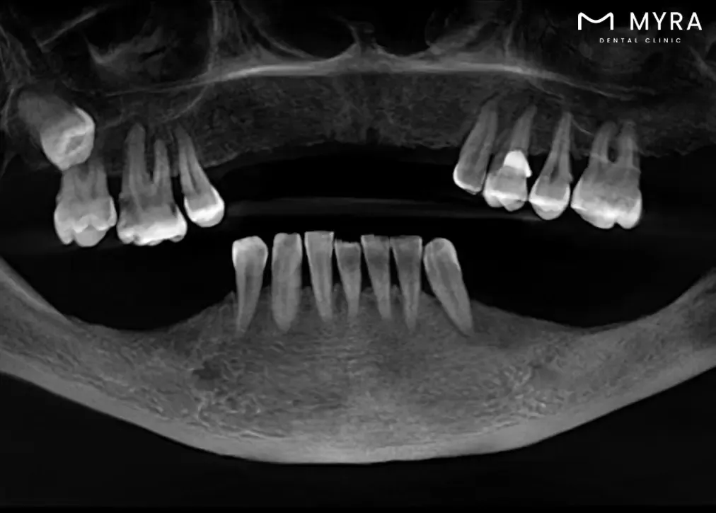
What does a 3D tomography X ray show?
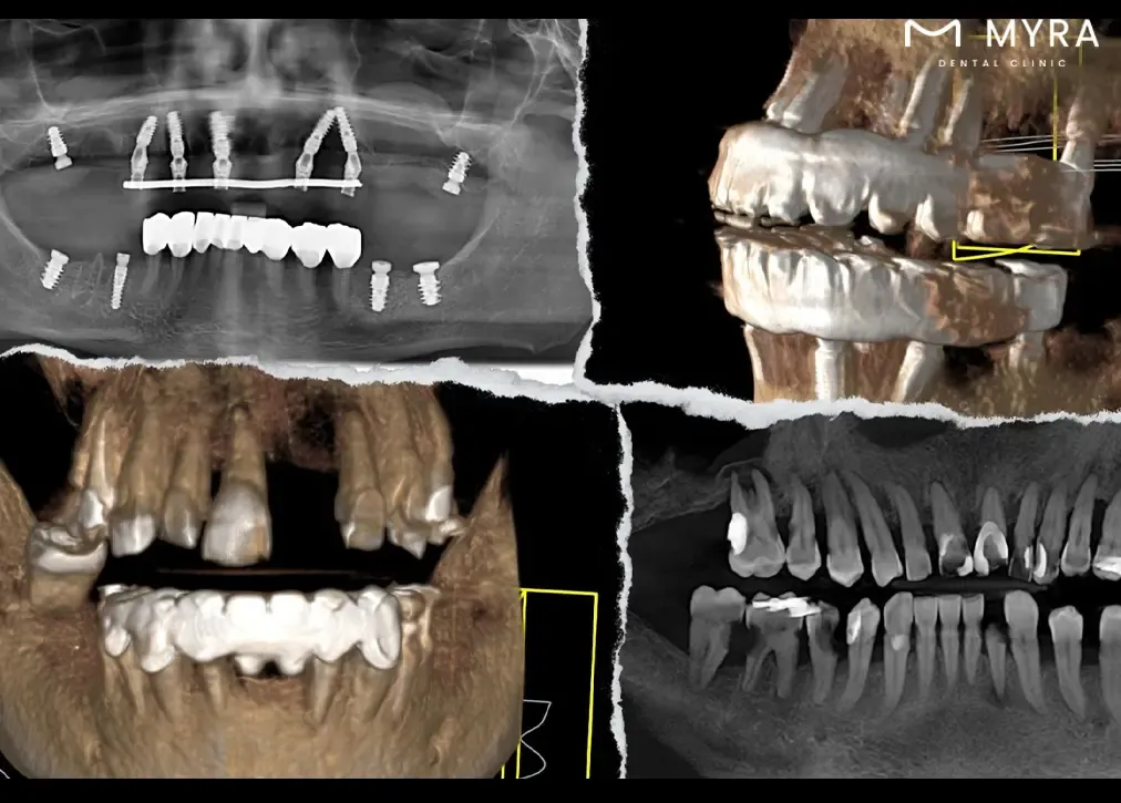
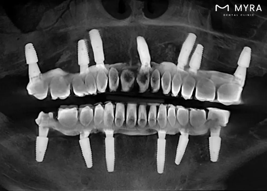
A 3D tomography X-ray shows a comprehensive view of the entire mouth, offering detailed insights into dental and jaw structures. The imaging method helps doctors better understand dental or jaw problems and come up with effective treatments by showing internal structures, bone density, soft tissues, and nerve distances. It makes it easier to diagnose dental problems such as cavities, buried teeth, fractures, bone defects, and other issues. It serves as a valuable tool in treatment planning, especially for dental implant placement, orthodontic procedures , and various oral health assessments , ultimately enhancing the success of treatments.
What is the role of Sirona 3D Dental Tomography in dental practises?
The role of Sirona 3D Dental Tomography in dental practises is to serve as the pinnacle device for advanced diagnosis and treatment in dental radiology. The technology’s significance lies in its ability to provide high-end images of the cranio-maxillofacial imaging area, offering a detailed map of the oral and dental structures. The device's Adaptive Focal Positioning (AFP) function ensures high clarity in imaging, allowing for precise analysis of multiple layers in exposures.
The Sirona 3D Dental Tomography is very useful because it combines different tomography layers into a single image that ensures all parts are visible, from the tooth's crown to the area between the teeth. The technology goes beyond traditional 2D X-rays, allowing for the viewing of hidden details that are crucial for diagnosis, treatment planning, and follow-up stages of oral and dental health.
The Sirona 3D Dental Tomography is frequently used for various purposes, including determining bruises, planning and following up on implant surgeries, detecting bone damage in advanced gingival diseases , and assessing joint disorders. The device is instrumental in cases involving cysts and tumours related to teeth and bones, determining the position of buried teeth before surgical procedures, and monitoring children's dental development and growth.
The Sirona 3D Dental Tomography's capacity to generate precise 3D images that aid in the diagnosis of conditions such as buried teeth, abnormal canal numbers, fractures, and cyst spread further emphasises its role. Its benefits extend to providing better image quality and higher accuracy, offering clear views of bone and soft tissues with a low radiation dose, and ensuring a quick and painless solution for patients.
What is the working principle of Sirona 3D Dental Tomography?
The working principles of Sirona 3D Dental Tomography are listed below.
- Adaptive Focal Positioning (AFP): AFP function ensures high clarity in imaging by analysing multiple layer exposures. It controls each region, selects optimal tomography layers, and transforms them into a new image. The adaptive nature of its function allows for customization based on specific diagnostic needs.
- Two-Dimensional Imaging: A two-way image is taken, providing a baseline for subsequent 3D imaging. The step involves capturing standard X-ray images that lay the foundation for more detailed scans.
- Rotational Scanning: The device rotates 180 degrees around the patient's head during the scanning process. The rotational scanning captures detailed images from different angles, contributing to the three-dimensional reconstruction.
- Computer Software Processing: Advanced computer software processes the captured images, creating sections as thin as two-tenths of a millimetre. The sections are compiled to generate the final 3D images.
- Volumetric Imaging: The device creates a volumetric image of the skull, allowing for a comprehensive examination of the bone structure. The three-dimensional view enhances the diagnostic capabilities of dental tomography.
- Focused Image Reconstruction: The adaptive nature of the focal positioning function ensures that each element, from the apex of the tooth to the incisal region, is perfectly in focus. The focused reconstruction contributes to the clarity and accuracy of the images.
- Conical Beam CT Technology: Sirona 3D Dental Tomography employs conical beam CT technology, which focuses X-rays at a specific point, enabling the imaging of small areas with high precision. The technology is a key part of getting more accurate diagnoses.
In what cases is Sirona 3D Dental Tomography used?
Sirona 3D Dental Tomography is used in the cases listed below.
- Implant Surgery Planning: Sirona 3D Dental Tomography is utilised to assess the bone structures of the jaw, allowing for precise planning of implant surgeries. The detailed images aid in determining the optimal placement of dental implants.
- Orthodontic Treatment Planning: The technology helps in evaluating the relationship between jaws and teeth in orthodontics. It assists orthodontists in planning and implementing effective treatment methods for malocclusions and misalignments.
- Root Canal Treatment: Sirona 3D Dental Tomography is used to visualise the internal structures of teeth before and during root canal treatment. It aids in identifying root canal anatomy and abnormalities and determining the extent of treatment required.
- Detection of Bruises and Fractures: The technology is valuable in identifying dental and jaw fractures and in finding bruises in the bone structures. It is crucial for an accurate diagnosis and appropriate treatment.
- Gingival Diseases and Bone Damage: Sirona 3D Dental Tomography is employed in cases where gingival diseases are advanced, helping to detect bone damage associated with these conditions.
- Cysts and Tumours: The tomography is used to investigate and determine the presence of cysts and tumours related to teeth and bones. The detailed imaging allows for early diagnosis and appropriate treatment planning.
- Salivary Gland Diseases: Sirona 3D Dental Tomography aids in the assessment of salivary gland diseases, allowing for accurate diagnosis and treatment decisions.
- Monitoring Dental Development in Children: Tomography is used for monitoring the dental development and growth of children. The technology provides insights into the positioning of teeth and helps in planning appropriate interventions.
- Joint Disorders: Sirona 3D Dental Tomography is valuable in assessing joint disorders in the cranio-maxillofacial region.
- Determination of Tooth Position Before Surgery: The equipment is useful for pinpointing the exact location and relationship with anatomical areas in situations where impacted teeth or buried teeth require surgical intervention. Suspected Tooth and Jaw Fractures: The equipment is used in cases where there is suspicion of fractures in the teeth and jaw for the dentist to obtain detailed pictures for precise diagnosis.
- Suspected Tooth and Jaw Fractures: The equipment is used in cases where there is suspicion of fractures in the teeth and jaw for the dentist to obtain detailed pictures for precise diagnosis.
- Dental Treatment Planning and Follow-Up: Sirona 3D Dental Tomography is widely used in dental treatment planning and follow-up stages, contributing to the success of oral and dental health interventions.
How is Sirona 3D Dental Tomography different from traditional dental x-rays?
Sirona 3D Dental Tomography is different from traditional dental X-rays, which are listed below.
- Dimensionality: Traditional dental X-rays are two-dimensional, presenting a flat representation of teeth and surrounding structures. Sirona 3D Dental Tomography offers three-dimensional imaging, allowing a more thorough analysis of dental and jaw anatomy from various angles.
- Level of Detail: Traditional X-rays lack the depth and clarity necessary to visualise fine details and structures. Sirona 3D Dental Tomography provides high-resolution images, offering a more precise view of dental and jaw conditions, including bone structures and soft tissues.
- Diagnostic Capability: Traditional X-rays are limited in their diagnostic capabilities for detecting certain conditions, such as subtle bone defects or complex anatomical relationships. Sirona 3D Dental Tomography enhances diagnostic accuracy, making it more effective in identifying a broader range of dental issues, from root canal configurations to bone density measurements.
- Treatment Planning: Traditional X-rays require additional imaging methods for comprehensive treatment planning. Sirona 3D Dental Tomography facilitates comprehensive treatment planning within a single imaging session due to its ability to capture detailed three-dimensional data.
- Radiation Exposure: Traditional X-rays expose individuals to greater radiation doses. Sirona 3D Dental Tomography uses lower radiation doses, contributing to safer imaging practises.
What are the advantages of Sirona 3D Dental Tomography for the patient?
The advantages of Sirona 3D Dental Tomography for the patient are listed below.
- Better Diagnosis and Treatment Planning: Sirona 3D Dental Tomography provides high-quality, three-dimensional images of the entire mouth, offering a comprehensive view that aids in the accurate diagnosis of dental and jaw problems. Its enhanced visualisation contributes to more effective treatment planning.
- Detailed Imaging for Treatment Stages: The technology is frequently used in various treatment stages, including planning for implant surgery by advanced procedures, root canal treatment, and orthodontic interventions. The detailed imaging allows for precise assessments, contributing to successful outcomes in these procedures.
- Reduced Radiation Exposure: Sirona 3D Dental Tomography involves lower radiation doses compared to traditional X-rays. It is advantageous for patients, as it minimises potential risks associated with radiation exposure while providing detailed and valuable imaging information.
- Early Detection of Issues: The technology enables the early detection of various dental problems, such as tooth decay, bone defects, and abnormalities. Early diagnosis allows for timely intervention and preventive measures, contributing to better oral health outcomes.
- Improved Visualisation of Hidden Details: Sirona 3D Dental Tomography excels at displaying hidden details that are not visible in two-dimensional X-rays. Its capability is especially valuable for identifying subtle issues in the bone structure, soft tissues, and tooth anatomy.
- Enhanced Treatment Success: The detailed images generated by Sirona 3D Dental Tomography contribute to a higher treatment success rate. Dental procedures, implant placements, and orthodontic treatments are all improved by technology, which increases the accuracy and effectiveness of treatment.
- Efficient Planning for Implant Surgery: The technology is particularly useful in planning and guiding implant surgeries. It allows for a thorough evaluation of the jawbone structure, facilitating precise implant placement and increasing the likelihood of successful outcomes.
- Comprehensive Examination of Oral Health: Sirona 3D Dental Tomography covers the entire mouth in a single image, offering a holistic view of oral health. Its comprehensive examination helps in identifying a range of issues, from bruising and root canal problems to joint disorders and salivary gland diseases.
- Useful in Orthodontic Treatment: The technology plays a crucial role in orthodontic treatment by aiding in the assessment of relationships between jaws and teeth. It assists in determining treatment methods and monitoring the development and growth of children's dental structures.
- Quick and Painless Procedure: The procedure for Sirona 3D Dental Tomography is relatively quick, usually taking 10 to 40 seconds. It provides a painless solution for patients, offering convenience without compromising the quality of imaging.
What are the disadvantages of Sirona 3D Dental Tomography for the patient?
The disadvantages of Sirona 3D Dental Tomography for the patient are listed below.
- Financial Considerations: Advanced dental imaging technologies, including Sirona 3D Dental Tomography, entail higher costs compared to traditional X-rays, posing potential financial concerns for certain patients.
- Limited Accessibility: The availability of Sirona 3D Dental Tomography varies, and not all dental practises possess advanced technology. Patients in specific locations or with limited access to specialised dental facilities find it challenging to undergo the specific imaging procedure.
- Requirement for Specialised Equipment: The use of Sirona 3D Dental Tomography necessitates specialised equipment, which is not universally available in every dental clinic. Patients need to visit specific facilities equipped with the necessary technology, potentially causing inconvenience.
- Pregnancy and Radiation Apprehensions: Pregnant women continue to have concerns about radiation exposure despite the safety of dental X-rays, including Sirona 3D Dental Tomography. Some pregnant women prefer not to undergo radiation-related procedures, even though the doses are minimised.
- Dependence on Technician Proficiency: The quality of imaging and the success of Sirona 3D Dental Tomography rely on the skills and experience of the technician operating the equipment. Discrepancies in technician expertise impact the effectiveness of the procedure.
- Complementary Use with Other Diagnostic Tools: Indications that traditional dental radiography or tomography continues to be necessary indicate that Sirona 3D Dental Tomography does not entirely replace all diagnostic tools. It leads to a combination of different imaging methods, potentially elevating radiation exposure.
- Limitations in Soft Tissue Visualization: Sirona 3D Dental Tomography has limitations in providing detailed images of soft tissues and contrast variations while excelling in visualising hard tissues. Some diagnostic scenarios require additional imaging methods for a more comprehensive assessment.
- Risk of Blurred Images with Movement: The information suggests that patient movement during the imaging process poses a risk of blurring the images. Patients are advised to remain still during the procedure to ensure the clarity of the tomographic images.
- Environmental Implications: Traditional X-ray films used in dental radiography are mentioned to have certain environmental impacts due to the chemicals involved in the film development process. Sirona 3D Dental Tomography, being a digital method, addresses the mentioned environmental concern.
How does Sirona 3D Dental Tomography help dentists?
Sirona 3D Dental Tomography helps dentists in several ways, which are listed below.
- Improved Diagnostic Accuracy: Sirona 3D Dental Tomography presents detailed three-dimensional images, allowing dentists to visualise dental and jaw structures with higher precision. It leads to more accurate diagnoses of dental conditions and abnormalities.
- Treatment Planning: Dentists utilise the comprehensive images generated by Sirona 3D Dental Tomography for precise treatment planning. The detailed view of teeth, roots, and surrounding tissues aids in formulating effective and tailored treatment strategies.
- Risk Assessment: The three-dimensional imaging capability allows dentists to assess the risk factors associated with various dental procedures, especially surgeries and implant placements. It assists in identifying potential challenges and planning interventions accordingly.
- Quick Diagnoses: Sirona 3D Dental Tomography facilitates quick and efficient diagnoses due to its ability to capture a comprehensive view in a single scan. It expedites the diagnostic process, allowing for prompt initiation of necessary treatments.
- Patient Education: The detailed visualisations provided by Sirona 3D Dental Tomography serve as valuable educational tools for patients. Dentists use these images to explain dental conditions, treatment plans, and potential risks, enhancing patient understanding and involvement in their own care.
- Reduced Radiation Exposure: Sirona 3D Dental Tomography involves lower radiation doses compared to traditional radiography. It ensures that patients receive the necessary diagnostic information with minimal radiation exposure, aligning with principles of patient safety.
How common is Sirona 3D Dental Tomography in dentistry?
Sirona 3D Dental Tomography has become common in dentistry because of its notable upswing among contemporary practitioners in the field, reflecting a growing inclination among dentists worldwide to embrace the advantages and innovations offered by the technology. Sirona, as a distinguished manufacturer, has played a significant role in driving the widespread adoption of 3D Dental Tomography throughout the dental industry.
One key indicator of the technology's prevalence is the consistent evolution of Sirona's imaging systems, which embody state-of-the-art advancements. The dental field tends to gravitate towards technologies that bring heightened capabilities, precision, and diagnostic accuracy. The reputation of Sirona contributes substantially to the broad acceptance of its cutting-edge equipment among dental professionals.
Professional endorsements and recommendations further underscore the prevalence of Sirona 3D Dental Tomography. Technology's role in dental practises is to enhance diagnostic accuracy and facilitate effective treatment planning. Dentists, influenced by professional commendations, are more likely to integrate Sirona's 3D Dental Tomography into their practises.
The rapidly increasing need for informed patients seeking advanced diagnostic methods has become another driving force behind the widespread adoption of sophisticated imaging technologies. Dentists increasingly recognise the need to incorporate Sirona 3D Dental Tomography into their practises in response to patient expectations and the pursuit of optimal care.
The emphasis on patient safety, particularly in terms of minimising radiation exposure, contributes to the widespread acceptance of Sirona's systems. The mention of reduced radiation concerns aligns with the industry's commitment to safety measures, making Sirona 3D Dental Tomography a preferred choice among dental professionals.
How does the Sirona 3D Dental Tomography procedure work?
The Sirona 3D Dental Tomography procedure works on a principle that involves capturing full three-dimensional images of the maxillofacial and dental structures. The process utilises advanced imaging technology to provide comprehensive views of the teeth, jawbone, and surrounding tissues.
The procedure begins with the patient being positioned appropriately, usually with the jaw placed in the designated area of the imaging device. The Sirona 3D Dental Tomography device rotates around the patient's head, capturing multiple X-ray images from various angles. A precise and three-dimensional picture of the maxillofacial and oral structures is captured using the rotational movement.
Sirona 3D Dental Tomography allows dentists to gain a more in-depth understanding of dental and jaw-related issues. The three-dimensional images enable precise and accurate diagnoses of conditions such as tooth decay, buried or impacted teeth, fractures resulting from trauma, bone defects, and the periodic assessment of implants.
The procedure aids in treatment planning by offering a higher level of detail and accuracy. Dentists configure root canals, measure root canal dimensions, and conduct detailed assessments of bone density, sinuses, and joints. The technology helps in the planning of dental implant procedures, offering a three-dimensional evaluation of the jawbone structure.
The Sirona 3D Dental Tomography procedure is designed to contribute to higher treatment success rates by providing dentists with advanced diagnostic tools. Dental professionals are able to improve their diagnostic capabilities and create more effective and personalised treatment plans with the ability to provide detailed and precise images.
How long does it take to get results from Sirona 3D Dental Tomography?
The waiting time to get results from Sirona 3D Dental Tomography is short as the result is available after the imaging session, offering a quick turnaround for the dentist and the patient.
The efficient nature of the Sirona 3D Dental Tomography process allows for rapid image acquisition. The entire imaging session takes between 5 to10 minutes, including the preparation time. The dentist or radiology technician promptly accesses the detailed three-dimensional images once the imaging is complete.
The speed at which the results are obtained is advantageous for treatment planning and decision-making. The 3D tomography results give dentists the ability to quickly analyse the images, make accurate diagnoses, and develop customised treatment plans.
The quick turnaround time enhances the patient experience. Patients receive timely feedback on their oral health status, allowing for efficient communication between the dental professional and the individual undergoing the procedure.
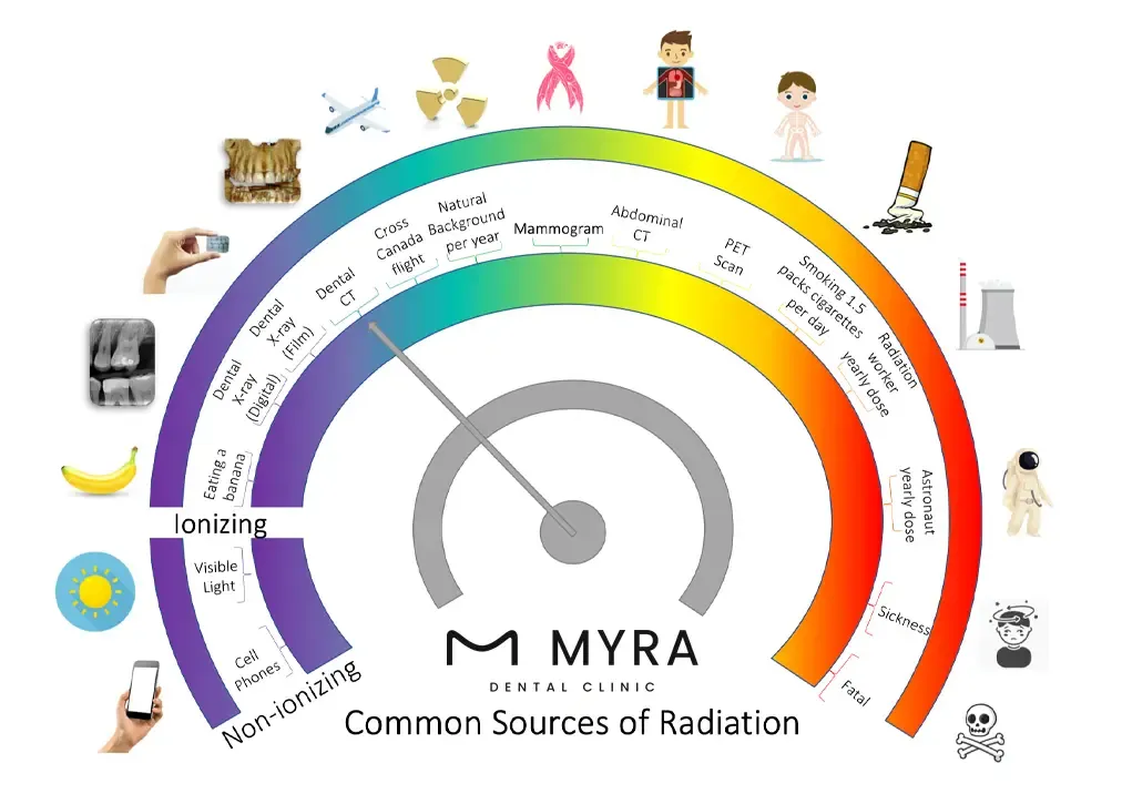
How low are the radiation levels for Sirona 3D Dental Tomography?
The radiation levels for Sirona 3D Dental Tomography are notably low, ensuring patient safety while providing high-quality diagnostic images. Sirona 3D Dental Tomography employs advanced technology that significantly reduces radiation exposure compared to traditional imaging methods.
Sirona 3D Dental Tomography uses lower radiation doses compared to conventional X-rays, making it a safer option for patients. It is achieved through innovative imaging techniques, such as cone-beam computed tomography (CBCT), which allows for precise and targeted imaging of the oral and maxillofacial structures with minimal radiation.
The specific radiation dose varies depending on the imaging parameters selected for each case, but in general, Sirona 3D Dental Tomography aims to minimise radiation exposure while maximising diagnostic accuracy. The technology is designed to focus on the area of interest, reducing unnecessary radiation to surrounding tissues.
The digital nature of Sirona 3D Dental Tomography contributes to dose reduction. Digital imaging systems are more efficient in capturing and processing images, requiring lower radiation doses to achieve the desired diagnostic quality.
Which patients are not suitable for the Sirona 3D Dental Tomography application?
Patients who are not suitable for the Sirona Dental Tomography application are listed below.
- Pregnant women: Women who are pregnant must limit their radiation exposure to the greatest extent feasible, especially during the first trimester. Sirona 3D Dental Tomography uses just a small amount of radiation but pregnant patients are urged to think about getting another kind of imaging or delaying the treatment.
- Children: Careful deliberation is required before using Sirona 3D Dental Tomography on children, even though it turns out to be necessary in some paediatric circumstances. Dentists weigh the benefits against the potential risks, especially considering the increased sensitivity of developing tissues to radiation.
- Radiation Sensitivity: Individuals with a history of radiation therapy or with known sensitivity to radiation must discuss the potential risks and benefits with their healthcare providers. The decision to proceed with Sirona 3D Dental Tomography must be made based on a thorough assessment of individual health conditions.
- Financial Considerations: Financial constraints limit access to Sirona 3D Dental Tomography. Patients without sufficient health insurance or financial means must look into other diagnostic alternatives if the technology turns out to be more expensive than conventional imaging techniques.
- Metallic Implants or Devices: Patients with certain types of metallic implants or devices, such as cochlear implants or certain prosthetic devices, are susceptible to imaging issues that affect the quality of the results. Alternative imaging methods are recommended in such cases.
Are there any risks or side effects of Sirona 3D Dental Tomography?
Yes, there are risks or side effects of Sirona 3D Dental Tomography similar to any medical imaging technique. The benefits outweigh the potential risks, and the procedure is generally considered safe when used with caution.
There is exposure to ionising radiation during Sirona 3D Dental Tomography. However, the radiation levels are relatively low, and advancements in technology have allowed for reduced doses compared to traditional radiographic methods. The benefits of precise diagnosis and treatment planning frequently outweigh the minimal risk that radiation poses.
Pregnant individuals must exercise caution, especially during the first trimester. Pregnant women must choose other imaging techniques that do not use ionising radiation to reduce the danger to their unborn child, even though the dose is small.
Metallic implants or dental restorations cause artefacts in the images, affecting their clarity. It limits the usefulness of the images in some cases.
People must tell their dentists about any preexisting conditions, including pregnancy, before having Sirona 3D Dental Tomography. It allows for a personalised assessment of the potential risks and benefits based on individual health considerations. Dentists take appropriate measures to minimise risks and ensure the safety of the patient.
How are the images obtained with Sirona 3D Dental Tomography evaluated?
The evaluation of images obtained with Sirona 3D Dental Tomography is listed below.
- Teeth Examination: Identification of cavities, fractures, or abnormalities in individual teeth. Assessment of the roots and nerve canals for any signs of pathology.
- Bone Structure Analysis: Evaluation of jawbones for density, defects, and anomalies. Detection of bone abnormalities or irregularities.
- Soft Tissue Assessment: Examination of gums and oral mucosa for abnormalities or signs of infection. Visualisation of soft tissue structures in the oral cavity.
- Implant Planning: Precise analysis of bone density and structure for optimal implant placement. Identification of potential challenges or anatomical variations.
- Pathological Conditions: Detection and analysis of pathological conditions such as cysts or tumours. Comprehensive assessment of abnormalities in the oral and maxillofacial region.
- Treatment Planning: Tailoring effective and targeted treatment plans based on the comprehensive information provided by the images. Planning for procedures such as extractions, root canals, and orthodontic treatments.
How much does Sirona 3D Dental Tomography cost?
The cost of Sirona 3D Dental Tomography is £150 but other prices vary depending on several factors, and it typically encompasses various aspects of the imaging procedure. Patients interested in cutting-edge dental imaging technology are advised to familiarise themselves with the factors that go into determining the final price.
The cost of Sirona 3D Dental Tomography surgery encompasses the expenses associated with acquiring high-quality images using advanced technology. It includes the use of the Sirona 3D imaging system, the expertise of the dental professionals performing the procedure, and the interpretation of the results. The surgery cost includes additional fees related to the imaging facility and post-surgery consultations.
The total cost is based on the type and quality of materials used in the Sirona 3D Dental Tomography process. High-end imaging systems and advanced radiographic materials contribute to increased expenses. The specific area or body parts being scanned impact the cost. Full-mouth scans or scans covering a larger anatomical region, incur higher charges compared to more localised scans.
The cost reflects the comprehensive nature of the dental services provided if the Sirona 3D Dental Tomography is part of a larger treatment plan that includes other procedures. The facility where the Sirona 3D Dental Tomography is performed charges additional fees. State-of-the-art facilities with advanced technology and amenities have higher associated costs. The experience and expertise of the dental professionals conducting and interpreting the procedure influence the cost. Highly skilled professionals charge higher fees for their services.
Patients must consult their dentist or oral health professional regarding the procedure's specifics and any related expenses. Many dental offices offer consultation appointments where patients inquire about the cost, payment options, and potential insurance coverage for Sirona 3D Dental Tomography.
How much does a Sirona 3D Dental Tomography Cost in Turkey?
The cost of a Sirona 3D Dental Tomograph in Turkey is £48. The quality and type of materials used during the Sirona 3D Dental Tomography process significantly sway the costs. Choosing cutting-edge imaging technology and superior radiographic materials contributes to variations in total expenses.
The body parts or areas targeted during the scan impact the total cost. Prices vary depending on the scope of the scan, which ranges from a thorough full-mouth scan to a targeted analysis of particular areas.
The cost reflects the comprehensive nature of the dental services provided if the Sirona 3D Dental Tomography is part of a broader treatment plan involving additional dental procedures. Consider the integration of various treatments in understanding the total expense.
Navigating the financial aspects of Sirona 3D Dental Tomography requires open communication with dental professionals. Discussing payment options and potential insurance coverage ensures a transparent understanding of the financial implications associated with its advanced dental imaging procedure in the Turkish context. Dental treatment prices in Turkey, including advanced procedures such as 3D dental tomography, are renowned for their cost-effectiveness and exceptional value.
How much does a Sirona 3D Dental Tomography Cost in Antalya?
The cost of Sirona 3D Dental Tomography in Antalya is £13. The price point at Myra Dental Center Turkey covers the Sirona 3D Dental Tomography procedure, providing patients with a high-quality imaging experience. The fee includes the utilisation of advanced materials, ensuring a precise and detailed examination.
Several factors contribute to the cost of Sirona 3D Dental Tomography. The type and quality of materials employed during the procedure play a significant role, with a focus on delivering top-notch imaging results. The specific body parts or areas being scanned influence costs, tailoring the procedure to individual needs. Integrating tomography into a larger treatment plan encompassing multiple dental procedures increases the cost because of the comprehensive nature of the dental services supplied.
Myra Dental Center Turkey stands for its commitment to providing high-quality dental services and to making advanced procedures such as Sirona 3D Dental Tomography accessible and affordable. Patients expect top-tier imaging technology and professional expertise at a cost that aligns with their budget, reinforcing the clinic's dedication to delivering exceptional dental care in Antalya.
What are the limitations of 3D Tomography X-ray?
The limitations of 3D Tomography X-ray are listed below.
- Exposure to Radiation: Ionising radiation is nevertheless exposed when using 3D tomography X-rays, despite efforts to minimise it. It is essential to consider the potential risks associated with radiation, even though the doses are generally low.
- Cost Considerations: The implementation of 3D Tomography X-ray technology comes with initial setup costs for dental practises. The costs are a limiting factor for smaller clinics or patients with budget constraints.
- Not Ideal for Routine Use: Common X-rays are enough for simple exams and regular dental checkups. 3D Tomography X-rays are powerful tools but they are not necessary for every case, and their use must be judicious.
- Not Suitable for All Patients: Certain patients, such as pregnant women, need to avoid 3D tomography X-rays due to concerns about radiation exposure. Alternative imaging methods are preferred in such cases.
- Overlapping Structures: Structures in the oral cavity overlap in the 3D images in some instances, making it challenging to interpret certain details accurately.
- Limited Availability: 3D tomography X-ray devices are becoming more common, although not every dentist's office has one on hand. The limitation impacts the availability of the procedure for some patients.
3D Tomography X ray Frequently Asked Questions
Share your phone number with us, we'll call you!