The gingival margin, frequently referred to as the marginal gingiva or the marginal ridge of the tooth, is critical to maintaining dental health. The gingival margin forms a protective barrier surrounding the teeth, shielding the underlying structures from dangerous bacteria and external particles. It is located near the gum line and leads to a variety of dental health complications when weakened.
Infections in the gingival margin are characterised by redness, swelling, pain, and bleeding. Illnesses such as gingivitis and periodontitis have the power to change the gingival margin, causing inflammation and damage to the tooth-supporting tissues. Understanding the importance of the gingival margin and recognising the signs of infection or common diseases is critical for maintaining good dental hygiene.
What is Gingival Margins?
The term "gingival margins," alternatively referred to as the marginal ridge of the tooth, denotes the demarcation line separating the gums and the teeth. The visible boundaries of the gingiva encircle safeguard the dentition, establishing a barrier between the tooth structure and the underlying supportive tissues. The gingival margins comprise the marginal gingiva, which constitutes the outermost layer of the gingival tissue. The narrow strip of tissue serves as a protective barrier, effectively obstructing the entry of bacteria, food particles, and other detrimental substances into the oral cavity, thereby preventing their infiltration into deeper layers.
The gingival margins play a crucial role in preserving oral health by anchoring the teeth and safeguarding the underlying structures, including the periodontal ligaments and the alveolar bone. Ensuring the well-being of the gingival margins is of utmost importance, necessitating adherence to appropriate oral hygiene protocols, periodic dental examinations, and timely intervention in the presence of any indications of infection or pathology.

Where is Gingival Margins located?
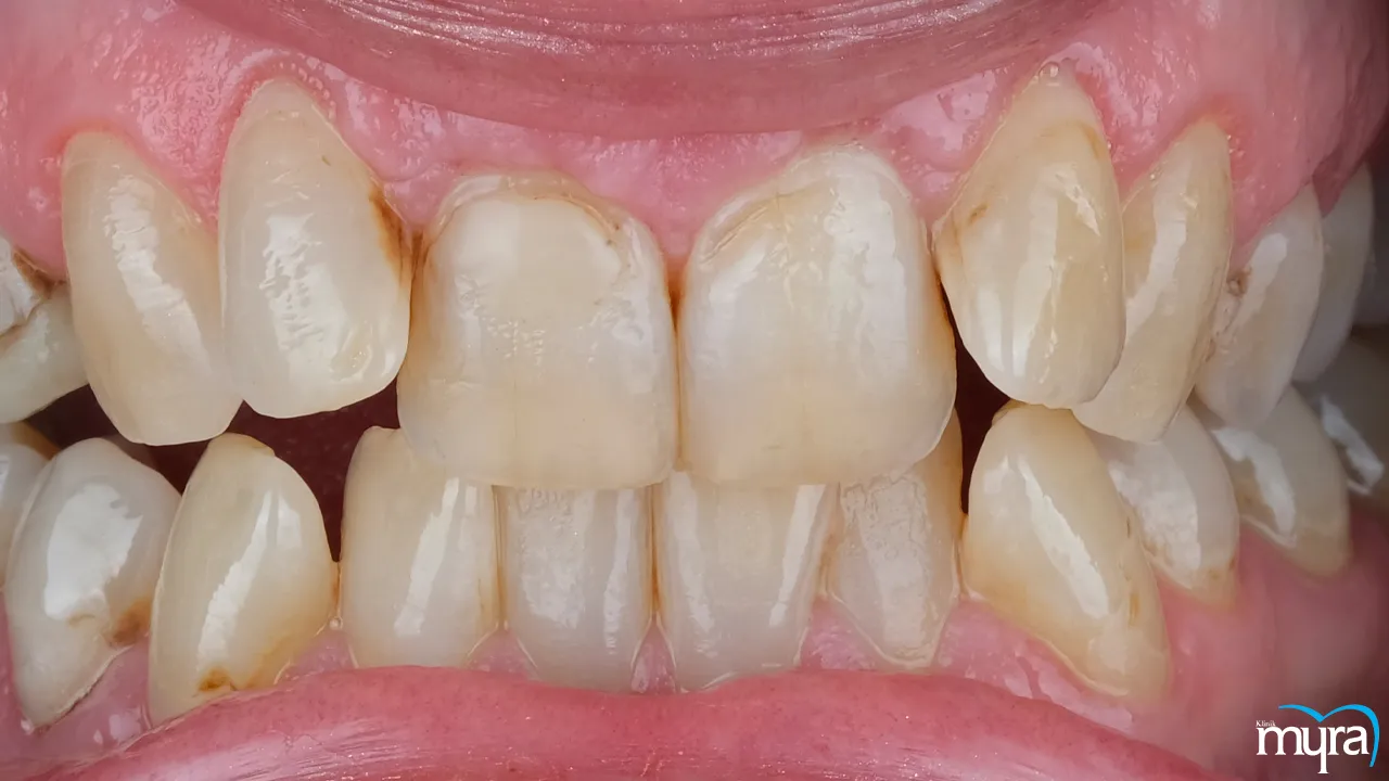
Gingival margins are found at the junction of the gums and the teeth. Gingival margins are most commonly found near the visible edge of the gum tissue where it meets the tooth surface. They are the lines where the pinkish gum tissue meets the white enamel of the tooth.
The gingival margins surround each tooth, forming a protective barrier that isolates the cervical area of the tooth. The gingival margins go all the way around the tooth, encompassing both the front and back surfaces. The gingival borders must fit tightly on the tooth surface in a healthy mouth, forming a tight seal. The tight seal prevents dangerous bacteria, food particles, and other debris from entering the gap between the teeth and the gums.
It is essential to remember that the gingival borders' position varies slightly depending on factors including tooth form, gum thickness, and individual anatomy. The gingival margins on the tooth tend to be somewhat higher or lower in some people, but they usually stay within a few millimetres of the tooth surface. Regular dental check-ups and expert cleanings are helpful in maintaining healthy and properly positioned gingival margins.
What is the Function of Gingival Margins?
The gingival margins serve multiple essential roles in the preservation of oral health. Gingival margins function as a defensive barrier, effectively sealing the gingival crevice where the periodontal tissues interface with the tooth structure. The presence of such barriers effectively hinders the ingress of detrimental bacteria, food debris, and other irritants into the interdental space, thereby mitigating the chances of infection and subsequent harm.
The gingival margins serve the important function of providing attachment and stability by securely anchoring the gingiva to the teeth. Gingival margins maintain the position of the gums and inhibit their recession or detachment. The gingival margins are essential in promoting oral hygiene as they guide toothbrush bristles and dental floss, ensuring that they follow the natural contour of the teeth. Such a technique facilitates the efficient elimination of plaque, bacteria, and food particles from the surfaces of the teeth and the gum line.
The copious vascularisation in the gingival margins is important in maintaining the well-being of the gum tissue by facilitating the delivery of oxygen, nutrients, and immune cells necessary for the processes of healing and immune defence. The nerve endings located in the gingival margins enable individuals to perceive sensory stimuli such as pressure, touch, temperature, and pain, thereby facilitating the identification of oral health concerns such as gum inflammation, recession, or tooth sensitivity.
Gingival margins play a crucial role in safeguarding, stabilising, promoting cleanliness, facilitating blood supply, and offering sensory input. Gingival margins underscore the importance of appropriate maintenance and monitoring practices to uphold optimal oral well-being.

What Are the Symptoms of Infection of Gingival Margins?
Listed below are the common symptoms that indicate an infection of the gingival margins.
- Redness and Swelling: The gingival margins frequently exhibit signs of infection, characterised by a reddened and swollen appearance. The gingiva is prone to symptoms associated with inflammation or oedema.
- Gum Bleeding: The occurrence of bleeding during oral hygiene activities such as brushing, flossing, or even without any external stimulation is a prevalent indication of gum infection. The gingival margins that are affected by infection exhibit a heightened susceptibility to bleeding as a result of heightened sensitivity and inflammation.
- Gum Recession: Infections have the ability to induce gingival recession or the detachment of gum tissue from the teeth, thereby exposing the underlying root surfaces. The current recession results in dental hypersensitivity and aesthetic considerations.
- Pain or Discomfort: The gingival margins that are inflamed exhibit tenderness upon palpation or result in discomfort, particularly during mastication or the application of pressure to the affected region.
- Bad Breath (Halitosis): Chronic halitosis serves as an indicator of an underlying infection within the gingival margins. The bacteria that are linked to gum infections have the ability to generate malodorous compounds.
- Gum Pockets: The development of deep spaces or pockets between the gums and teeth occurs during the advanced stages of gum infection. Such pockets serve as reservoirs for bacteria, rendering them resistant to conventional oral hygiene practices such as brushing and flossing.
- Pus or Abscess Formation: The gingival margins are prone to the formation of pus or abscesses in instances of severe infection. Such localised areas of infection necessitate prompt dental intervention.
- Loose or Shifting Teeth: There is an opportunity for the gradual deterioration of the supporting structures as the gum infections progress, resulting in tooth mobility or displacement. Such a condition is more pronounced in cases of advanced periodontal disease.
- Changes in Bite or Tooth Alignment: Severe periodontal diseases have the capacity to impact dental occlusion and disrupt the harmonious relationship between the maxilla and mandible. Changes in bite or tooth alignment occur over time.
1. Redness and Swelling
The presence of redness and swelling in the gingival margins commonly signifies the presence of inflammation in the periodontal tissues. The gingival margins show visible signs of erythema and enlargement or oedema as a response to irritation or infection.
Redness and swelling do not always suggest a severe infection, but they serve as early indicators of gum disease or gingivitis. Gingivitis has the tendency to advance into periodontitis if left untreated. Periodontitis is a more advanced manifestation of gum disease that leads to substantial harm to the gums and supporting structures.
Enhancement of oral hygiene practices is frequently employed to address the occurrence of gingival margin redness and swelling. The implementation of consistent oral hygiene practices, such as regular brushing and flossing, in conjunction with periodic professional dental cleanings, has been shown to effectively eliminate plaque and bacteria.
Such practices result in a reduction of inflammation and facilitate the process of gum tissue restoration. It is advisable to pursue supplementary interventions such as scaling and root planing or antimicrobial rinses in cases of heightened severity.
2. Gum Bleeding
Gum bleeding is characterised by the occurrence of blood during oral hygiene practices such as brushing, flossing, or even without any external stimulation. Gingival inflammation, resulting in heightened sensitivity, leads to increased susceptibility to bleeding at the gingival margins.
Infrequent and minor bleeding during oral hygiene is not an immediate cause for alarm, but continuous or excessive bleeding is indicative of an underlying condition such as gum disease or more advanced infection.
Addressing the underlying cause of gum bleeding is of paramount importance in order to mitigate subsequent harm to the gums and tooth loss. The management of gingival bleeding entails the identification and resolution of the root cause, frequently attributed to periodontal disease. Interventions for enhancing oral hygiene include various strategies such as refining oral hygiene practices, employing antibacterial mouthwashes, and seeking professional dental cleanings. Pursuing more comprehensive periodontal treatments such as scaling and root planing or gum surgery is done during severe cases.
3. Gum Recession
Gum recession is characterised by the detachment of gum tissue from the teeth, resulting in the exposure of the root surfaces. Gum recession is caused by gum disease, excessive brushing force, malocclusion, and various other contributing factors. The gum recession issue is of considerable importance due to its consequences, including heightened tooth sensitivity, heightened susceptibility to decay, and aesthetic implications. Tooth roots that are exposed are prone to damage and eventually exacerbate the advancement of periodontal disease.
The management of gingival recession is contingent upon the extent of the condition and its root aetiology. The implementation of enhanced oral hygiene practices, utilisation of a toothbrush with soft bristles, and the identification and resolution of underlying factors such as tooth misalignment impede or cease the progression of recession. Surgical interventions, such as gum grafting, become imperative in order to reinstate gum tissue and provide coverage for roots that have become exposed during severe cases of infection.
4. Pain or Discomfort
The sensation of pain or discomfort experienced in the gingival margins varies in intensity, ranging from mild sensitivity to more severe sensations such as sharp or throbbing pain. Persistent pain or discomfort occurs during the act of chewing, dental hygiene practices, or even during periods of inactivity. The presence of chronic pain or discomfort in the gingival margins indicates the presence of an underlying infection, periodontal disease, or other oral health conditions. Such an observation suggests an advanced periodontal disease or the formation of an abscess.
The management of pain or discomfort in the gingival margins is contingent upon the etiological factors. The treatment options for dental issues involve professional dental cleaning, administration of medication for infection or pain relief, or more advanced interventions such as root canal therapy or periodontal procedures. The identification and treatment of the underlying cause is of paramount importance in alleviating pain and mitigating the risk of subsequent complications.
5. Bad Breath (Halitosis)
Halitosis, commonly known as bad breath, is characterised by the presence of a consistently unpleasant odour emanating from the oral cavity. The condition happens due to the accumulation of bacteria and subsequent infection in the gingival margins. The occurrence of bad breath is attributed to multiple factors, but the presence of persistent halitosis serves as an indicator of an underlying condition such as gum disease or an infection in the gingival margins. Addressing the underlying cause is of paramount importance in order to mitigate the occurrence of subsequent oral health complications.
The management of halitosis entails the identification and resolution of its root cause. Interventions to enhance oral health encompass various strategies such as optimising oral hygiene practices, managing gum disease or infections, employing antibacterial mouthwashes, and addressing underlying factors such as xerostomia or dietary habits. The provision of professional dental care facilitates the identification and management of the fundamental factors contributing to halitosis.
6. Gum Pockets
Gum pockets are anatomical recesses that develop within the periodontal tissues, specifically between the gingiva and the teeth. Periapical abscesses arise from the detachment of gum tissue, thereby creating a conducive environment for the entrapment of bacteria and debris. Gum pockets give rise to subsequent infection and consequential harm. They are indicative of the presence of advanced periodontal disease or periodontitis. Such variables are of significant concern as they have been found to be associated with the occurrence of bone loss, tooth mobility, and an elevated likelihood of tooth loss if not promptly addressed.
The management of gum pockets typically necessitates professional dental intervention. The dental procedure known as scaling and root planing is conducted with the purpose of eliminating the accumulation of plaque and tartar below the gum line, as well as achieving a smoother texture on the surfaces of the tooth roots. It facilitates the reattachment of the gums and subsequent reduction in pocket depth. Surgical interventions are required on certain occasions to further decrease the pocket depth and facilitate gum reattachment.
7. Pus or Abscess Formation
The occurrence of pus or abscess formation signifies the existence of an infection in the gingival margins or adjacent tissues. Pus or abscess formation is distinguished by the presence of an inflamed and tender region containing purulent fluid. It is indicative of a severe infection that necessitates prompt medical intervention. Its occurrence suggests the presence of an advanced periodontal disease or a localised gingival abscess.
The course of treatment encompasses the evacuation of the abscess to eliminate the purulent material, as well as the administration of antibiotics to manage the infection. It is imperative to address the root cause, such as periodontal disease or dental infection, in order to effectively prevent the recurrence of abscesses. The provision of professional dental care is imperative in order to accurately diagnose and effectively treat abscesses.
8. Loose or Shifting Teeth
The condition of loose or shifting teeth pertains to teeth that have acquired mobility or are undergoing positional alterations. Loose or shifting teeth is frequently correlated with the progression of severe periodontal disease and subsequent deterioration of the underlying bone structure. Its presence is indicative of substantial impairment to the underlying supportive structures, encompassing the gingival margins, periodontal ligaments, and osseous tissues. It is a serious condition that results in the loss of teeth if left untreated.
The selection of treatment modalities is contingent upon the aetiology and extent of the condition. Periodontal therapies, such as deep cleaning, gum surgery, or tooth stabilisation techniques, are employed to mitigate increased tooth mobility and enhance oral health. The extraction of a tooth and subsequent replacement with dental implants or alternative prosthetic solutions are deemed necessary in severe conditions.
9. Changes in Bite or Tooth Alignment
Changes in bite or tooth alignment pertain to modifications in the occlusion of the upper and lower dentition. Periodontal disease and its consequential bone resorption have a detrimental impact on dental stability and bite alignment. Alterations in bite or tooth alignment serve as indicators of advanced periodontal disease and harm to the supporting structures. Such a condition has the capacity to give rise to functional impairments and additional medical complexities if not given the appropriate intervention immediately.
The treatment regimen includes interventions aimed at managing gum disease and mitigating bone loss in order to stabilise the teeth and reinstate optimal bite alignment. Common interventions for addressing bite and tooth alignment issues involve a range of dental procedures, such as periodontal treatments, orthodontic interventions, or restorative measures, such as crowns or bridges. The timely initiation of intervention plays a pivotal role in mitigating subsequent bite-related complications and maintaining optimal oral functionality.

Can dentures cause gum recession or gingival margin changes?
Yes, dentures cause gum recession or gingival margin changes. Dentures are a form of dental prosthetics that are designed to be removable and serve the purpose of replacing teeth that are missing, thereby restoring both the functionality and visual appeal of the oral cavity. The use of dentures results in various consequences on the gums and gingival margins as time progresses. The application of force and the resulting resistance between dentures and the gingival tissues lead to various adverse effects, such as irritation, inflammation, and harm to the gingival margins.
The absence of natural teeth contributes to bone resorption, which in turn induces alterations in the morphology and dimensions of the underlying jawbone. Such a phenomenon has the power to impact the suitability and steadiness of dentures, thereby resulting in heightened pressure exerted on the gingival tissues and the occurrence of gum recession. The presence of ill-fitting dentures ultimately exacerbates gum recession due to the trauma inflicted upon the gums.
Inadequate oral hygiene practices and insufficient denture maintenance give rise to bacterial or fungal infections, exacerbating gum inflammation and precipitating gum recession. It is advisable for individuals to adhere to a regimen of effective oral hygiene, remove their dentures during the night to provide respite for the gums, and maintain regular appointments with a dentist for assessments, modifications, and appropriate denture maintenance. Such practices are important to mitigate the chances of gum-related complications linked to dental dentures.
Can gingival margins affect implant healing and integration?
Yes, gingival margins affect implant healing and integration. The gingival margins, or gums, are crucial to the process for a number of reasons. Gingival margins offer a barrier of defence around the dental implant, protecting the supporting tissues during the healing process. It assists in fostering the best environment for healing and guards against bacterial invasion, which hinders implant integration.
The gingival margins surrounding an implant must be healthy and stable for it to be successful over time. The gum tissue surrounding the implant gets inflamed or infected, which impedes healing and hinders the integration of the implant with the surrounding bone. The associated gingiva, or the tough, keratinised gum tissue bound to the underlying bone, is crucial in terms of its thickness and quality. Enough connected gingiva maintains the health of the peri-implant tissues and helps shield the implant from mechanical pressures. It makes it easier to maintain good dental hygiene.
The final restoration's aesthetics and natural appearance are significantly influenced by the positioning and shapes of the gingival borders. Gingival margins that are appropriately made and sculpted help create a beautiful smile and a successful prosthesis. The state and health of the gingival margins have a significant impact on how quickly dental implants heal and integrate. The success and long-term stability of dental implants are mainly dependent on maintaining a healthy soft tissue environment, protecting the peri-implant mucosa, guaranteeing adequate connected gingiva, and carefully controlling the location and shapes of the gingival borders.
How to prevent common gingival margin diseases?
The maintenance of optimal oral health necessitates the prevention of common gingival margin diseases. It is imperative to consistently engage in the practise of maintaining proper oral hygiene. Brushing one's teeth a minimum of two times per day with a toothpaste containing fluoride and a toothbrush with soft bristles effectively eliminates plaque and food debris. The gentle cleaning of the tongue and gums and the daily practice of flossing or utilising interdental brushes help mitigate the risk of developing gum disease.
Routine dental examinations and cleanings conducted by dental professionals, such as dentists or dental hygienists, hold equal significance in the detection of initial indications of gingival margin diseases and the administration of prompt therapeutic interventions. The adoption of tobacco avoidance practices and limiting alcohol consumption have been found to have a substantial impact on the reduction of gum disease susceptibility. A diet that is well-balanced and contains an abundance of essential vitamins and minerals plays a crucial role in promoting optimal immune system functioning and maintaining healthy gum tissue.
Engaging in physical exercise, practising meditation, or engaging in social interactions with close companions are effective strategies for enhancing the resilience of the immune system in coping with stress. The inclusion of an antimicrobial mouthwash in one's oral hygiene regimen has been shown to effectively decrease the presence of bacteria in anatomical regions that are difficult to access.
Maintaining proper hydration through regular consumption of ample amounts of water throughout the day facilitates the removal of food particles and bacteria. Individuals effectively uphold the health of their gingival margins and mitigate the likelihood of prevalent gingival margin diseases by adhering to such preventive measures and availing themselves of professional dental care.
What are common gingival margin diseases and conditions?
Listed below are the common gingival margin diseases and conditions.
- Periodontitis: Periodontitis is a highly consequential manifestation of periodontal disease, distinguished by inflammation and infection that explicitly impacts the gingival margins and supportive anatomical components of the teeth, encompassing the periodontal ligaments and alveolar bone. It leads to the occurrence of gum recession, tooth mobility, and ultimately tooth loss if left untreated.
- Gingival Hyperplasia: Gingival hyperplasia is a pathological condition characterised by excessive enlargement and overgrowth of the gingival margins. The aetiology of the condition is attributed to various factors, including the administration of certain medications, alterations in hormonal levels, or an inherent genetic susceptibility. The particular condition leads to challenges in upholding oral hygiene practices and increases the susceptibility of the gums to infection.
- Gum Resorption: Gum resorption is a pathological condition characterised by the progressive loss of gingival tissue surrounding the teeth, typically attributed to factors such as traumatic events, inflammatory processes, or mechanical forces. Gum resorption results in the uncovering of tooth roots, thereby elevating the susceptibility to tooth sensitivity and dental caries.
- Gingival Abscess: A gingival abscess refers to a circumscribed and distressing enlargement of the gingival tissue, frequently resulting from a bacterial infection. The occurrence of such a condition arises due to the entrapment of food particles within the interdental spaces or the presence of advanced periodontal disease.
- Oral Lichen Planus: Oral lichen planus is a persistent inflammatory disorder that has a risk of impacting the gingival margins and various other oral tissues. The manifestation is characterised by the presence of white, lace-like patches or areas that are red and swollen. The precise aetiology remains uncertain, but it is hypothesised that the condition is associated with an immune system response.
- Oral Thrush: Oral thrush, referred to as oral candidiasis, is a fungal infection that manifests within the oral cavity, specifically affecting the gingival margins. The condition appears as depigmented patches on the gingiva and lingual mucosa and is prevalent among individuals with compromised immune systems or those undergoing specific pharmacological treatments.
- Gum Tumours: Gingival tumours encompass a spectrum of growths that appear as either benign or malignant tumours within the gingival margins or other gum tissues. The timely identification and comprehensive assessment are imperative for accurate diagnosis and effective intervention.
1. Periodontitis
Periodontitis is a severe form of gum disease that destroys the gums and the periodontal ligaments and alveolar bone that keep teeth in place. Plaque and calculus (tartar) formed by microorganisms on and around teeth cause chronic irritation of the gums. Gum recession causes spaces between teeth and gums, which bacteria easily colonise as the infection worsens. The periodontal ligaments and bones that support the teeth deteriorate over time due to the body's immune reaction to the bacteria and their metabolites. It leads to gum recession, loose teeth, and eventually tooth loss if not treated immediately.
Periodontitis is not always avoidable, but the risk is significantly decreased by maintaining a regular routine of proper dental hygiene. Plaque and tartar buildup, which is associated with gum disease, is easily reduced with regular brushing, flossing, and expert dental cleanings. Avoiding tobacco usage and eating a healthy, well-balanced diet is additionally helpful in maintaining good oral health.
There is currently no cure for periodontitis, but the disease is managed, and its progression is prevented with the help of medication. Treatment and diagnosis must begin immediately. Deep cleaning methods called scale and root planing are used in the treatment to get rid of plaque and tartar beneath the gum line. Surgery is required in more severe situations to treat deep pockets and stimulate bone and gum regeneration. Some gum recession and bone loss are permanent, but teeth are saved with the help of treatment that stops the problem from worsening.
2. Gingival Hyperplasia
Overgrowth and enlargement of the gum tissue is the hallmark of gingival hyperplasia, frequently referred to as gingival overgrowth. Medications such as anticonvulsants and calcium channel blockers, hormonal shifts, and genetic predisposition are common triggers. Overgrowth makes it harder to practise good oral hygiene, which in turn raises the risk of gum disease and other problems related to the mouth.
The development of gingival hyperplasia is difficult to prevent in cases where it is associated with medication use or underlying medical conditions. Good oral hygiene and routine dental checkups are crucial for early diagnosing and treating such a condition.
Gingival hyperplasia is a chronic condition and is not always curable, especially if it is brought on by medication. Careful oral hygiene, regular dental cleanings, and therapy help lessen its severity and keep it under control. Overgrowth is occasionally managed by discussing a drug adjustment or switch with the prescriber.
3. Gum resorption
Gum resorption occurs when the gum tissue that surrounds the teeth recedes for any number of reasons, such as infection, inflammation, or physical pressure. Roots of teeth become more sensitive and susceptible to decay if gums begin to recede.
Gum resorption is not always entirely preventable because of factors such as genetics and trauma. The likelihood of gum resorption is lowered by practising diligent oral hygiene, not using too much force when brushing, and seeking immediate treatment for any dental injuries.
Gum resorption varies in severity, depending on the underlying cause. Gum resorption stabilises or improves with proper oral hygiene and care. More comprehensive therapies, such as gum grafting, are necessary in more severe cases to rebuild gum tissue and hide exposed tooth roots.
4. Gingival abscess
A gingival abscess refers to a circumscribed and distressing enlargement of the gingival tissue commonly attributed to a bacterial pathogenic invasion. Periodontitis occurs as a result of bacterial entrapment in the interdental spaces or as a consequence of severe periodontal disease.
The prevention of gingival abscesses entails the maintenance of optimal oral hygiene practices and the timely pursuit of dental treatment upon the emergence of any indications of gum inflammation or infection. Regular dental examinations play a crucial role in the early detection and management of common dental problems, thereby preventing their progression into abscesses.
Gingival abscesses are amenable to treatment and are frequently resolved through appropriate drainage and administration of antibiotics. The dental professional has the option to perform an incision and drainage procedure on the abscess, which facilitates the release of pus and relieves the associated pressure. A prescription for antibiotics is then provided afterwards in order to manage the infection. It is imperative to address the root cause, such as the presence of advanced periodontal disease, in order to effectively prevent the recurrence of abscesses.
5. Oral lichen planus
Oral lichen planus is a persistent inflammatory disorder that impacts the gingival margins and various other oral tissues. Oral lichen planus manifests as patches resembling lace that are white in colour or as areas that are red and inflamed. Its exact origin remains uncertain, but it is hypothesised that the condition is associated with an immune system response.
The precise aetiology of oral lichen planus remains inadequately explained, thereby limiting the feasibility of complete prevention. Adhering to proper oral hygiene practises and seeking professional dental treatment effectively mitigate the condition and detect any complications.
Oral lichen planus is a persistent condition for which a definitive cure has not yet been identified. The primary objective of treatment is to effectively address symptoms and mitigate the presence of inflammation. Topical corticosteroids or other medications are frequently prescribed by dental professionals in order to mitigate discomfort and facilitate the process of wound healing. Regular follow-up appointments play a crucial role in the monitoring of the patient's condition and the timely addressing of any alterations or concerns that arise.
6. Oral thrush
Oral thrush, commonly referred to as oral candidiasis, is a fungal infection that manifests within the oral cavity, specifically affecting the gingival margins. Oral thrush manifests as depigmented patches on the gingiva and lingual surface and is prevalent among individuals with compromised immune function or those undergoing specific pharmacological treatments.
The prevention of oral thrush is achieved through the maintenance of optimal oral hygiene practices and the promotion of a robust immune system. The risk of thrush is mitigated by minimising the overuse of antibiotics and effectively managing underlying medical conditions that compromise the immune system.
Oral thrush is normally effectively managed through the administration of antifungal medications as prescribed by a healthcare practitioner. The implementation of an efficacious treatment regimen successfully eradicates the infection and mitigates its associated symptoms. It is imperative to recognise and tackle any fundamental factors that contribute to the occurrence of thrush in order to prevent its recurrence.
7. Gum tumours
Gingival tumours encompass a spectrum of growths that manifest as either benign or malignant tumours in the gingival margins or other gum tissues. The timely identification and comprehensive assessment of individuals are imperative in order to establish accurate diagnoses and administer suitable interventions.
The prevention of gum tumours entails the maintenance of optimal oral health and adherence to routine dental examinations. The timely identification of any abnormal alterations or proliferations in the gingival tissue facilitates early detection and enhances the efficacy of therapeutic interventions.
The successful treatment of gum tumours is contingent upon several factors, including the classification of the tumour as either benign or malignant, as well as its stage and specific anatomical site. Benign tumours are characterised by their non-malignant nature and frequently exhibit a favourable prognosis, amenable to surgical intervention for removal.
Malignant neoplasms necessitate more comprehensive therapeutic interventions, including surgical procedures, radiation therapy, and chemotherapy, contingent upon the severity of the condition. The timely identification and intervention of gum tumours are pivotal factors in the effective management of the condition.
How Is Gingival Margins Treated?
Gingival margin treatment must be tailored to the underlying cause of gum disease. Plaque and tartar buildup below the gum line is removed with professional dental cleaning techniques called scale and root planing to treat gingivitis, the most common form of gum disease. Antimicrobial mouthwashes are typically administered to prevent bacterial development and inflammation, and patients are trained on basic oral hygiene practices to maintain oral health at home.
Scaling and root planing are procedures used to clean below the gum line and smooth the root surfaces in order to encourage gum reattachment in cases of periodontitis, a more severe type of gum disease. Flap surgery and gum grafting are two surgical treatments that help minimise pocket depths, stimulate tissue regeneration, and restore gum health in more difficult situations.
Gingival hyperplasia requires adjustment or changing of certain medications if it is medication-induced. Patients are counselled on how to best practise meticulous oral hygiene to minimise plaque buildup along the gingival borders, in addition to frequent professional dental cleanings.
Gum grafting is necessary in extreme cases of gum resorption to replace gum tissue and cover exposed tooth roots, thereby preventing sensitivity and decay. Incision and drainage are common procedures for treating a gingival abscess, which relieves discomfort and pressure. Antibiotics are prescribed to control the infection and promote healing.
Chronic inflammation of the gingival margins and other oral tissues is caused by oral lichen planus, which is treated with topical corticosteroids to reduce inflammation and alleviate symptoms. Medication to inhibit the immune system is sometimes recommended when the immunological response becomes out of control. Oral thrush, a fungal infection of the mouth and its surrounding tissues is treatable with antifungal drugs.
Procedures such as a biopsy to collect a tissue sample for diagnosis and, in the case of benign tumours, surgical removal are part of the treatment plan for gum tumours. Radiation therapy and chemotherapy are sometimes used together to treat malignant tumours. A dentist or periodontist must be consulted for an accurate diagnosis and a tailored treatment plan for any problem affecting the gingival margin. Maintaining healthy gums and jawbones requires prompt diagnosis and treatment.

What Is The Natural Color Of Gingival Margins?
Gingival margins, known as gums, appear differently in colour depending on a person's race and skin tone. Pink or coral pink is the normal colour of healthy gingival margins. Variations in tooth colour, while not ideal, are unlikely to point to a problem with one's oral hygiene. It is common for some people to have gums that are a slightly different shade of pink than others.
Higher melanin levels are associated with darker skin, which is why people of African, Asian, or Middle Eastern heritage tend to have darker gingival margins. The term "gingival melanin pigmentation" is used to describe such a phenomenon, which is a perfectly normal variation that has no bearing on gum health.
Any change in colour, including redness, pallor, or bluish discolouration, must be taken seriously as it is an indication of a health problem or gingival disease. It is recommended to see a dentist or other trained oral health practitioner if there is concern about how the gums appear or feel.
What Shape Should Gingival Margins Be?
The ideal shape of gingival margins, or gums, must be curved gently and continuously around each tooth. The gingival margins must rise and fall gently between teeth, creating a scalloped or 'knife-edge' pattern. Such scalloped form is frequently compared to a mountain range's peaks and valleys.
Function and aesthetics equally depend on the gingival margins having the proper contour. The gums' scalloped form serves a practical purpose by shielding the tooth roots and underlying bone from wear and tear caused by chewing and talking. Properly shaped gingival margins contribute to an attractive smile because they frame the teeth in a way that seems natural and balanced.
The scalloped shape is considered ideal, but it is essential to remember that there are significant variations among people in the structure of the gingiva. Genetics and individual differences in gum tissue thickness give people more pronounced or less pronounced scalloped gingival edges. Such variations have little to no effect on oral health if the gums are otherwise healthy.
A periodontist or competent dentist conducts gum reshaping techniques such as gingivoplasty or gum contouring if there are aesthetic problems or gum irregularities to provide a more balanced and symmetrical appearance of the gingival borders.
A healthy and appropriately shaped gingival margin is an important part of oral health and helps to create an attractive smile, whether it is naturally scalloped or has modest individual variances. Regular dental check-ups and good oral hygiene practices are essential to maintaining healthy gingival margins and oral well-being.
What is the Aesthetic Importance of Gingival Margins?
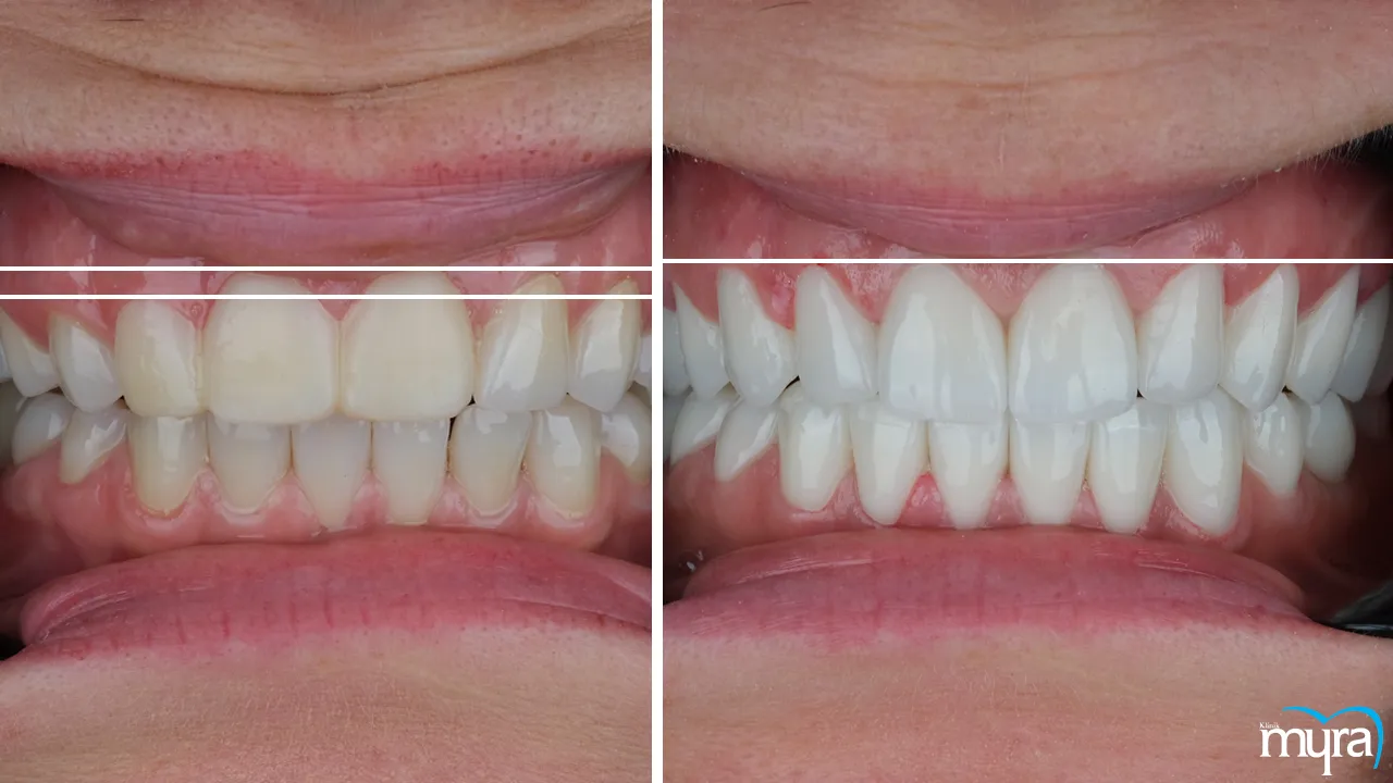
Gingival margins, or gums, play an essential function in the aesthetics of a person's smile and the balance of their face as a whole. The gingival margins are just as crucial to the aesthetic appeal of a smile as the teeth themselves. One key aspect is the smile line, formed by the upper edge of the upper front teeth when smiling, which must ideally follow a harmonious and smooth curve that complements the natural contours of the lips and face. Achieving the right balance between tooth display and gum display is vital, as excessive or uneven gum display, known as a "gummy smile," impacts the aesthetic value of a smile.
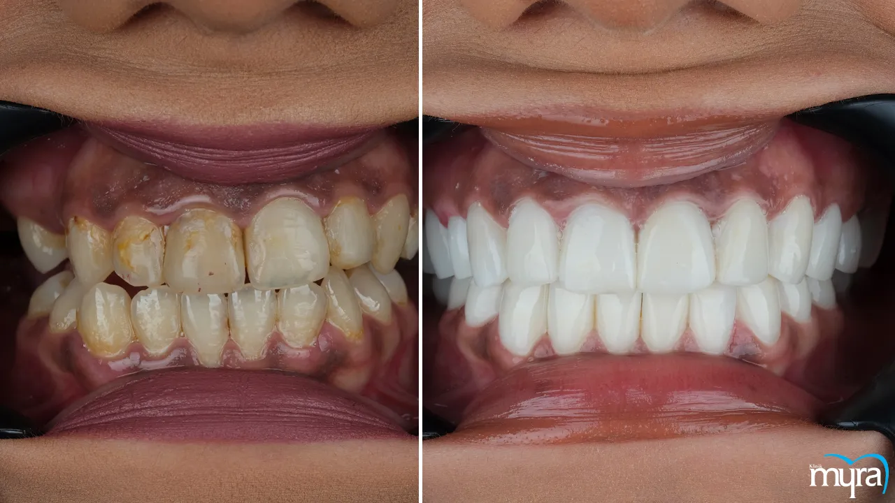
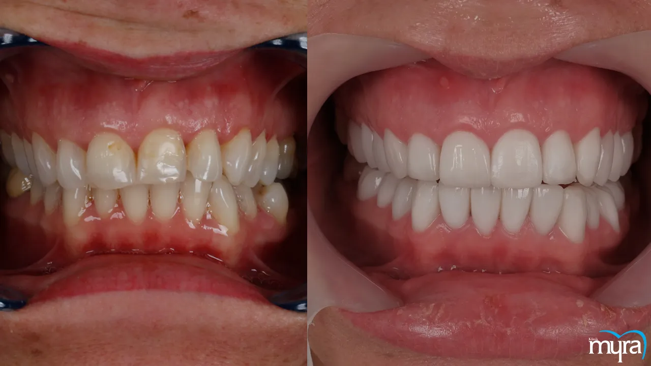
An aesthetically pleasant grin has a balanced gum-to-tooth ratio, which is achieved by having well-contoured gingival margins. Gingival margins affect how the teeth are perceived in terms of length and shape, improving their appearance. Gingival margins that are equally healthy and properly shaped serve as a frame for the teeth, drawing attention to their aesthetic value and providing a smooth transition between the original teeth and any dental restorations, such as veneers or crowns. An attractive grin has a significant psychological effect, increasing one's sense of self-worth and positively influencing one's interactions with others.
Gum contouring and reshaping operations are common in cosmetic dentistry and serve to improve the appearance of the gums and the smile as a whole. Gingival margins have a significant impact on the appearance of a person's smile and, by extension, on their confidence and sense of well-being. Regular dental check-ups and good oral hygiene are essential for preserving the aesthetic value of the gingival margins and maintaining gum health.
How does teeth whitening impact the appearance and color of the gingival margins?
The colour of the gums is not directly affected by teeth whitening, although the teeth themselves are brightened. The main goal of teeth whitening is to remove stains and discolourations from the tooth enamel, resulting in a more radiant and brighter smile. Whiter teeth stand out more against pink gums because of the stark contrast between the two. The enhanced contrast makes the gingival margins appear darker or more pigmented in comparison to the whiter teeth.
The gums' colour further plays a role in how white the teeth appear. The teeth next to them appear even whiter if the gums have a higher melanin pigmentation or are naturally darker. It's important to keep the aesthetic balance between the gums and teeth in mind to achieve a more harmonious appearance. Individuals with significant gum pigmentation ought to consider additional cosmetic procedures, such as gum depigmentation, to achieve a more uniform and balanced colour between the teeth and gums.
Consult a dental specialist first to examine the present colour of the teeth and gingival margins and to receive personalised advice to achieve the desired cosmetic result from teeth whitening. It is the best way to guarantee a successful teeth whitening treatment that is tailored to each person's distinctive smile and facial characteristics.


