Maxillary Central Incisors: Definition, Anatomy, and Common Procedures
HomeMyra BlogMaxillary Central Incisors: Definition, Anatomy, and Common Procedures
Dt. Erdem Çetin
Born in Stuttgart, Germany in 1979, Dr. Dt. Erdem Çetin began his education in Kırşehir and completed his schooling there, from primary school through to high school. He then successfully completed his university education at the Faculty of Dentistry at 19 Mayıs University in Samsun.
Maxillary central incisors are the two front teeth in the upper jaw positioned along the midline of the face. Maxillary central incisors serve a significant purpose in biting, breaking food, and creating the shape of the mouth, making them vital for function and appearance. The visible portion of the tooth above the gum line is called the crown, and the root, which is buried in the jawbone, makes up the maxillary central incisors anatomically. The crown has two mesial and distal sides and a flat incisal edge that helps with biting and chopping food. The incisors are fixed into the upper jawbone by a single root.
Several common procedures are involved with maxillary central incisors, including dental fillings, root canal treatment, dental crowns, orthodontic treatments, and cosmetic procedures. Dental fillings are frequently used to repair damaged tooth structures when decay causes a cavity. Dental amalgam or composite resin is applied similarly. Root canal therapy is often required to remove sick tissue, clean the root canal, and seal it to stop further infection if the pulp tissue inside the tooth becomes inflamed or infected.
Dental crowns are placed over the maxillary central incisors to restore their size, form, and strength in cases of severe decay, fractures, or aesthetic issues. Maxillary central incisors are crucial for tooth alignment and bite function. Braces or clear aligners are examples of orthodontic treatments that are often suggested to treat malocclusions involving them, overcrowding, or misalignment problems.
The maxillary central incisors are prominent teeth in the smile, thus for improved appearance and symmetry, cosmetic operations, including enamel shaping, dental veneers, or teeth whitening are frequently performed on them. Proper oral function and a confident smile depend on maintaining the health and aesthetic appeal of the maxillary central incisors. The preservation of the essential teeth depends upon routine dental examinations and good oral hygiene habits.
What are Maxillary Central Incisors?
Maxillary Central Incisors are the two front teeth in the upper jaw that are positioned in the centre of the face. The teeth provide support for dental health and appearance. Maxillary central incisors serve a crucial role in the early phases of digestion by allowing food to be bit and torn. Moreover, they aid in the production of sounds during the speech, particularly the ones involving the enunciation of certain consonants such as the letters “S” and “F.” The symmetry of the smile and the general attractiveness of the face are greatly enhanced by the noticeable visibility of those teeth when smiling. The maxillary central incisors are frequently the focus of different dental procedures because of their position and function. The procedures are intended to preserve the health, alignment, and aesthetic appeal of the teeth, ensuring optimal oral function and an attractive smile.
There are a few small variations between the permanent maxillary central incisor and the deciduous maxillary central incisor. Approximately 7-8 years old is when the permanent tooth erupts, replacing the deciduous tooth that first emerges in the mouth at 8–12 months of age. The permanent tooth has a longer length than width. The central incisors of the maxilla meet at the midline of the face. It is the mandibular central incisors that do it. The presence of an open bite or diastema depends on where the teeth are positioned. People differ in terms of the size, shape, and colour of their teeth, similar to any other. Teeth appearance is often impacted by systemic diseases, such as syphilis.
What is the anatomy of Maxillary Central Incisors?
The anatomy of Maxillary Central Incisors is listed below.
Crown: The portion of the tooth that is visible above the gum line is called the crown. Its incisal edge is somewhat flat, which facilitates biting and chopping food. The two sides of the crown that face neighbouring teeth are commonly known as the mesial and distal surfaces.
Incisal Edge: The maxillary central incisor's cutting edge is known as the incisal edge. It is the tooth's thin, sharp edge that is utilised to bite and shred food. The crown's cutting edge, facilitates food biting and ripping.
Root: The maxillary central incisor's root penetrates below the gum line to secure the tooth in the mandible. They usually have a single root that is thinner and shorter than the roots of the other teeth in the mouth.
Enamel: The toughest tissue in the human body, enamel, makes up the crown's outermost layer. Chewing wears down enamel, which acts as a barrier against acids and germs to stop deterioration.
Dentin: The majority of the tooth's structure is made up of dentin, a calcified tissue that is located beneath the enamel. Enamel is supported by dentin, which is a little bit softer than enamel.
Pulp Chamber: The pulp chamber is located in the centre of the crown and extends into the root. It is composed of connective tissue, blood vessels, and nerves. The pulp chamber is essential to the tooth's ability to sense and be fed.
Cementum: The thin layer of calcified tissue that surrounds the tooth root is called cementum. It acts as an anchor for the tooth about the surrounding bone and gives the periodontal ligaments places to connect. A strong tissue layer that covers the tooth root and helps hold it firmly in place in the jawbone.
Periodontal Ligament: The fibrous tissue that surrounds the root and connects the tooth to the surrounding bone is called the periodontal ligament. The periodontal ligament supports the tooth and permits a small amount of mobility within its socket.
Gingiva: The soft tissue that covers the jawbone and encircles the base of the tooth is known as the gingiva or gums. It offers a seal around the tooth to stop infection and aids in protecting the tooth's internal tissues. The gums surround the tooth, supporting and shielding the tooth's structure.
Incisal Fossa: The maxillary central incisor's cutting edge has a concavity called the incisal fossa. It aids in directing the opposing teeth's movement when biting and ripping.
1. Crown
The crown is essential for biting, ripping, and chewing food, which helps with the early phases of digestion. Cutting food particles is made effective by its flat incisal edge. The maxillary central incisors are quite noticeable when speaking or smiling, which greatly enhances the appearance of the crown. The visible part of a maxillary central incisor above the gum line is referred to as the crown. It performs several essential activities and is an essential part of the tooth's anatomy.
Self-confidence and social interactions are impacted by the crown's shape, size, and alignment, which affects how the face and smile are perceived. The crown guards against decay, infection, and mechanical injury to the tooth's underlying components, including the pulp chamber and dentin. Maintaining the health and integrity of the crown is crucial for optimal oral health and cosmetic appeal, as demonstrated by its relevance in the architecture of the maxillary central incisors.
2. Incisal Edge
The cutting edge that is sharp and situated at the apex of a maxillary central incisor is called the incisal edge. It is very important in tooth architecture and oral function. The incisal edge helps in biting and tearing food during the mastication process, which makes it easier for food particles to break down initially in preparation for digestion. Its sharpness makes it easy to cut through food, allowing for good chewing and swallowing. The incisal edge plays a significant part in the appearance of the smile and its practical use because it is easily noticeable when smiling or speaking.
An incisal edge that is well-defined and symmetrical improves the smile and teeth as a whole, boosting self-esteem and fostering pleasant social relationships. Keeping the incisal edge intact is crucial for dental health because any wear or damage on the edge affects the appearance and functionality of the tooth. Realising the role that the incisal edge plays in the architecture of the maxillary central incisors emphasises how important it is to maintain and properly care for the teeth to maintain both function and attractiveness.
3. Root
The root of a maxillary central incisor is the portion of the tooth that extends beneath the gumline and secures it to the jawbone. It is an essential part of the tooth's anatomy, giving its entire framework stability and support. Maintaining the tooth's position within the dental arch and making sure it is properly aligned with neighbouring teeth is crucial for efficient biting, chewing, and speaking. It is where the root comes into play. The pulp of the tooth, which receives essential nutrients and sensations from the blood vessels and nerve endings housed in the root, is where the tooth's pulp is located.
The root's neuronal and vascular network enables sensory perception, including the detection of pressure and temperature changes, enabling the body to react appropriately to outside stimuli. The root helps to maintain jawbone density and structure by conveying stresses generated during chewing and biting to the surrounding bone tissue, improving complete oral health and stability. Knowing the role that the root plays in the structure of the maxillary central incisors emphasises how important it is to preserve the root's integrity through good oral hygiene and dental care to guarantee stability and long-term oral function.
4. Enamel
Enamel is the outermost layer of a maxillary central incisor's crown and is known for its exceptional toughness and endurance. Enamel serves a crucial function in protecting the underlying tooth components from decay, erosion, and mechanical damage by acting as a screen against numerous environmental stimuli. Strong resistance to the corrosive effects of acidic meals and drinks and the abrasive pressures experienced during chewing and biting is provided by its thick crystalline structure. The smooth surface of enamel helps to avoid the accumulation of bacteria and plaque, which lowers the incidence of gum disease and dental caries.
Enamel's transparent quality, which lets the natural colour of the dentin underneath show through and gives the smile a glossy, lively appearance, greatly enhances its aesthetic appeal in addition to serving a protective purpose. Maintaining the strength of enamel through good oral hygiene practices, such as routine brushing, flossing, and dental check-ups, is crucial for guaranteeing the longevity and vitality of the maxillary central incisor and overall oral well-being because of its critical role in maintaining both oral health and aesthetics.
5. Dentin
The majority of the tooth structure is made up of dentin, which is an essential part of the anatomy of a maxillary central incisor. The pulp chamber lies behind the enamel layer and encircles the pulp chamber. It plays a variety of roles and is crucial to the general health and function of the tooth. Dentin acts as a buffer against outside forces and temperature fluctuations, giving the sensitive pulp tissue inside the tooth the vital protection and defence it needs. Dentin has intrinsic sensitivity, which allows it to sense pressure, temperature, and other stimuli. The sensitivity aids in controlling oral habits, such as biting and chewing.
Dentin helps to preserve the general health and integrity of the tooth by facilitating the right reaction to external stimuli and the transmission of sensory information to the pulp's nerve terminals. Dentin plays an important role in the tooth's cosmetic appearance because the colour and form of the tissue affect the enamel layer above the overall hue and translucency. The necessity of maintaining the health and integrity of dentin through regular dental care and strict oral hygiene habits is highlighted by the recognition of its relevance in the anatomy of the maxillary central incisors. It guarantees the best potential oral function and vitality.
6. Pulp Chamber
The pulp chamber is a central hollow found within the tooth that houses the intricate network of blood vessels, nerves, and connective tissue referred to as the dental pulp. It is an essential component of the anatomy of a maxillary central incisor. The pulp chamber is essential to the tooth's vitality, sensitivity, and overall health. The dental pulp is the lifeblood of the tooth, it provides the surrounding dentin and enamel with vital nutrients and oxygen, preserving the tooth's structure and sustenance.
The pulp chamber's sensory nerves permit the perception of a range of stimuli, including pain, pressure, and temperature, enabling the tooth to respond appropriately to outside influences and continue to operate. The pulp chamber's connective tissue promotes the durability and longevity of the tooth by helping dental tissues regenerate and mend after damage or infection. The maxillary central incisor's optimal function and vitality, and the general health of the mouth, depend heavily on preserving the pulp chamber's health and integrity through appropriate oral hygiene habits and prompt dental procedures.
7. Cementum
Cementum is a specialised calcified tissue that covers the root surface of a maxillary central incisor tooth and is an important part of its anatomy. The cementum, which acts as a contact between the tooth and the surrounding periodontal ligament, is vital for providing structural support and anchoring the tooth within the jawbone. Its main job is to make it easier for the periodontal ligament fibres to bind to the tooth and the surrounding alveolar bone. It keeps the tooth in its socket and allows for the small movement required for proper chewing and biting motions.
Cementum aids in shielding the root canal system and underlying dentin from microbial invasion and outside stimulation. It has some potential to regenerate and cure itself, which helps to preserve the stability and health of the periodontal system. The longevity and stability of the tooth within the oral cavity are dependent on maintaining the cementum's integrity, which is achieved by practising good oral hygiene and receiving regular dental treatment. It is made clear by understanding the cementum's significance in the anatomy of the maxillary central incisors.
8. Periodontal Ligaments
Periodontal ligaments (PDLs) are part of a maxillary central incisor's morphology and are fibrous connective tissues that enclose the tooth root and attach it within the alveolar bone socket. The stability and structure of the tooth within the dental arch are crucially maintained by these ligaments. PDLs serve as shock absorbers by equally distributing the stresses produced by biting and chewing across the tooth. It reduces stress on the surrounding bone tissue and stops excessive movement or displacement. PDLs allow for a small amount of tooth movement, which is necessary for orthodontic treatments and occlusal harmony maintenance.
PDLs facilitate accurate regulation of jaw motions during mastication and proprioceptive awareness by transferring tactile sensations to the surrounding tissues, thus contributing to sensory feedback. A PDL helps remodel alveolar bone in response to mechanical pressures and participates in tooth eruption, all of which contribute to the stability and health of the dentition. Maintaining the health and function of the teeth through good oral hygiene habits and routine dental check-ups is essential to achieving optimal oral health and function. It's true when considering the role that periodontal ligaments play in the structure of the maxillary central incisors.
9. Gingiva
The gingiva or gums, are an essential part of the structure of the maxillary central incisor. They maintain the stability and general health of the tooth by acting as a protective covering around it. The gingiva is important for dental health because it forms a seal around the base of the tooth, protecting the periodontal ligaments and alveolar bone from microbial invasion and mechanical damage. The gingiva distributes and absorbs forces applied during biting and chewing, acting as a cushion to lessen the chance of harm to the tooth and surrounding tissues.
A strong foundation for the tooth is provided by a healthy gingiva, which guarantees the tooth's stability inside the dental arch and encourages appropriate alignment and occlusion. The gingiva adds to the cosmetic look of the smile, framing the teeth and increasing the entire appeal aside from its functional significance. The integrity of the maxillary central incisor must be preserved, as general oral health and well-being, by maintaining the gingiva through regular oral hygiene habits, such as brushing, flossing, and routine dental check-ups.
10. Incisal Fossa
The concave depression on the cutting edge of the tooth's crown is known as the incisal fossa, and it is an anatomical feature of the maxillary central incisor. The tooth's appearance and functionality are greatly influenced by the special structure. The incisal fossa facilitates biting and ripping of food by creating a place for opposing teeth to occlude or come into contact with during chewing movements. It helps break down food particles for digestion by facilitating effective cutting and grinding processes. The incisal fossa lessens the chance of wear or injury to the incisal edge by helping to uniformly distribute the forces applied during biting and chewing across the tooth.
The incisal fossa's shape and contour contribute to the smile's entire aesthetic appeal, highlighting its symmetry and inherent attractiveness. The incisal fossa affects phonetics, specifically on how some sounds are pronounced that need the tongue to make contact with the teeth's incisal margins. Understanding the role of the incisal fossa in the anatomy of the maxillary central incisor highlights the relevance of the structure for oral function and aesthetics. It highlights the necessity of maintaining and providing appropriate dental care to protect the integrity of the structure and maintain the highest level of oral health and function.
What is the position of Maxillary Central Incisors?
The position of Maxillary Central Incisors is located in the maxilla, the bone that makes up the upper jaw, more precisely at the front portion. It is mesial, or near to the midline of the face, to the maxillary lateral incisor. The maxillary lateral incisors stand on either side of the teeth and are set exactly in the middle of the dental arch. They are positioned next to the face's midline, and when the mouth is closed, the ends of their crowns often match the upper lip's curvature. They are known as teeth numbers 8 and 9 in the Universal Numbering System, with tooth number 8 representing the right maxillary central incisor and tooth number 9 representing the left part.
The ideal position of maxillary central incisors is achieved when the GALL is in contact with the facial axis (FA) point. The goal anterior-limit line (GALL) is a line parallel to the frontal plane of the head that passes through the forehead's anterior-limit line (FALL) and the glabellar vertical line (GVL). The facial axis (FA) point is a clinical midpoint of the labial surface of the maxillary central incisors. The symmetry and attractiveness of the smile are greatly enhanced by its central placement inside the dental arch and alignment with the midline of the face.
What is the shape of Maxillary Central Incisors?
The shape of Maxillary Central Incisors is a trapezoidal or rectangular form, with unique features that add to their appearance and functionality. These teeth often have a larger mesiodistal dimension, compared to their height. The biting surface, known as the incisal edge, is straight and flat, forming a strong cutting edge that makes it easier to shred and bite food. The crown's distal and mesial surfaces curve inward toward the incisal border and have a small convexity.
Maxillary central incisors are characterised by a somewhat rounded or convex form that follows the lip's contour when viewed from a facial perspective. They are firmly anchored into the alveolar bone of the upper jaw by their typically straight, conical roots that taper towards the peak. The maxillary central incisors' design is designed to facilitate effective speaking, biting, and ripping actions while enhancing the appearance of a smile.
What is the function of Maxillary Central Incisors?
The maxillary central incisors perform several crucial functions that are necessary in terms of general health and oral health. These teeth are essential for biting and shredding food, which helps with the first phases of digestion. Food gets efficiently chopped into smaller, more digestible bits with the help of their sharp, incisive edges. The articulation of speech is made easier by the maxillary central incisors, which help generate sounds that are associated with the contact of the tongue with the front teeth.
The teeth are not just functional when it comes to eating and speaking, they contribute to the appearance of the smile. They enhance the appearance of the face because they are prominently positioned in the upper jaw and are noticeable when speaking, grinning, or laughing. The beauty of the smile is influenced by its symmetry, alignment, and colour, which affects social interactions and self-confidence. The maxillary central incisors support the lips and facial muscles, assisting in the maintenance of appropriate facial expressions and beauty. The maxillary central incisors play a critical role in maintaining both aesthetic harmony and oral function, underscoring the significance of their upkeep and health for complete dental health and well-being.
What are the common dental procedures for Maxillary Central Incisors?
The common dental procedures for Maxillary Central Incisors are listed below.
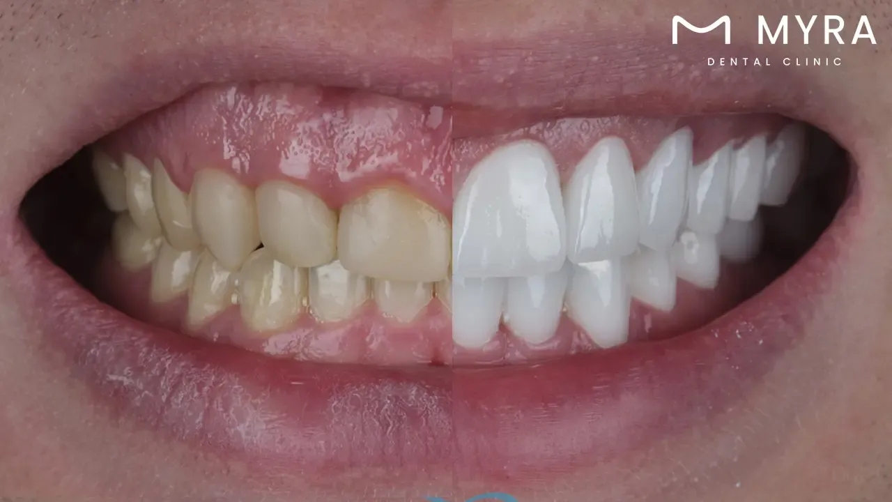
Dental Fillings: Dental fillings are commonly placed on damaged tooth structures when decay causes a cavity. Dental amalgam or composite resin is suitable for the application. The common methods for filling cavities and stopping additional decay are composite resin or dental amalgam materials.
Root Canal Treatment: Root canal therapy is often required to remove the infected tissue, clean the root canal, and seal it to stop further infection if the pulp tissue inside the maxillary central incisor becomes inflamed or infected. The goal of the treatment is to reduce pain and suffering while preserving the tooth from extraction.
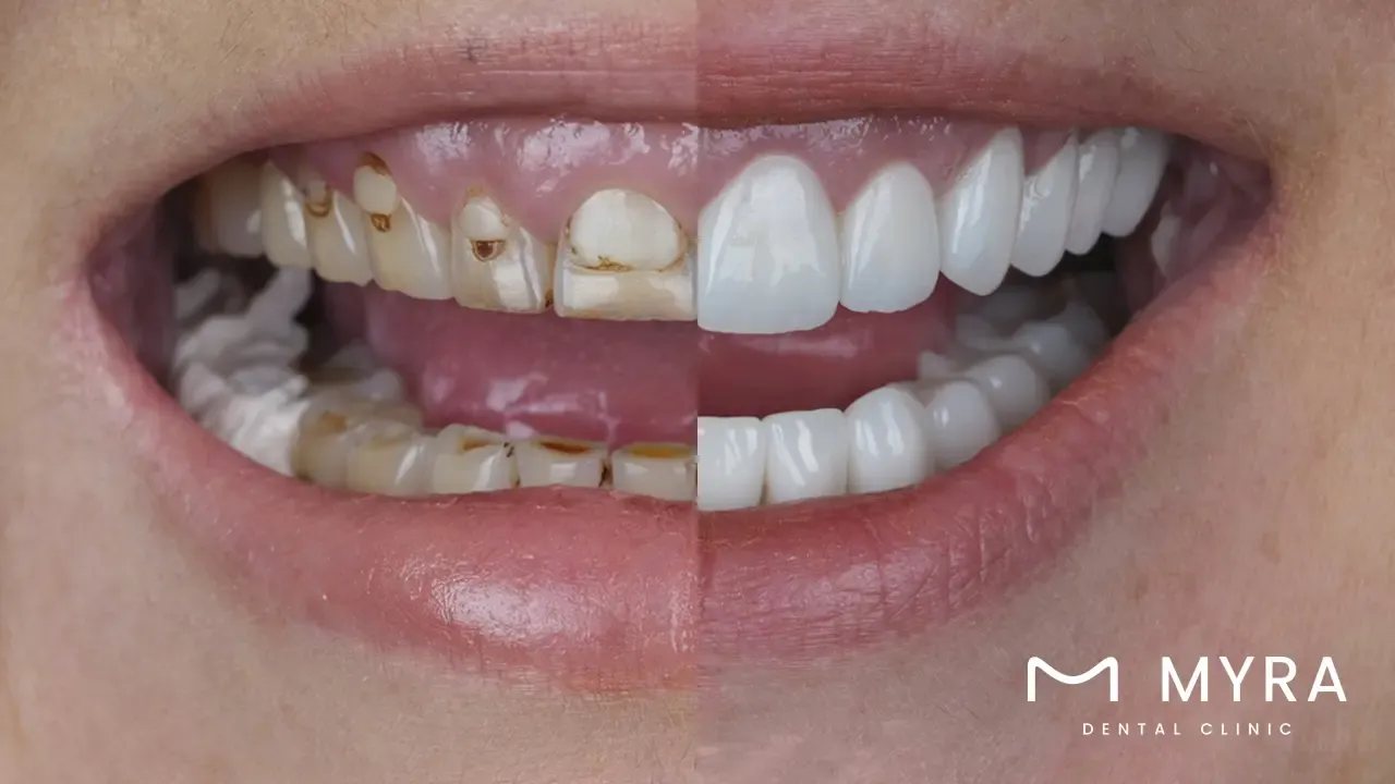
Dental Crowns: Dental crowns are usually placed over the maxillary central incisors to restore their size, form, and strength in cases of severe decay, fractures, or aesthetic issues. Crowns are prosthetic crowns, created to order, that completely encase the visible part of the tooth above the gum line, giving it a new appearance, size, strength, and form.
Dental Veneers: The front surfaces of the Maxillary Central Incisors are enhanced in look by dental veneers, which are thin shells composed of porcelain or composite resin that are attached to them. Veneers improve the appearance of the smile by fixing problems including discoloration, slight misalignment, chips, or cracks.
Orthodontic Treatment: Maxillary Central Incisors require orthodontic treatment, such as braces or clear aligners, to remedy misalignment, overcrowding, or malocclusion issues. The function and appearance of the smile are enhanced by orthodontic treatment, which aligns the teeth and bites appropriately.
Dental Implants: The procedure for dental implants is used as an alternative for lost maxillary central incisor teeth in situations when the tooth was extracted, lost to trauma, or decayed. A prosthetic crown that imitates the look and feel of a real tooth mounts on top of a titanium post that is surgically implanted into the jawbone to create a dental implant.
1. Dental Fillings
A dental filling is a popular dental technique used to cure cavities or tiny regions of tooth deterioration. An anaesthetic is administered initially to numb the area surrounding the damaged tooth so that each patient is comfortable throughout the procedure. A dental drill or alternative tools eliminate the decayed section of the tooth, leaving behind a clean and healthy tooth structure. The dentist uses an appropriate filling material to replace the cavity once the decay is gone to restore the tooth's strength, form, and function.
Dental amalgam is a long-lasting amalgam of silver, tin, copper, and mercury. A usual filling material is a composite resin, similarly shaped to real teeth, and attaches directly to the tooth surface. The filling component is meticulously moulded and polished to blend in with the existing tooth structure. Maxillary central incisors are teeth that are noticeable when smiling and are often filled because of their conspicuous position at the front of the mouth, which makes them prone to decay.
Several factors contribute to the prevalence of dental fillings for maxillary central incisors. The maxillary central incisors are more vulnerable to decay and damage than other teeth because they are the most noticeable teeth in the upper jaw. Many people prioritise their beauty because of their position, which makes them more apparent when they smile. The teeth are essential for chewing and biting, so any decay or damage has a big effect on oral health. Maxillary central incisors are susceptible to cavities because they are continually in contact with food particles and oral microorganisms, particularly if good oral hygiene habits are neglected.
Dental fillings are an efficient and minimally invasive treatment for treating cavities in maxillary central incisors, restoring the tooth's structure and strength while maintaining its natural appearance. Dentists stop potential decay and damage while maintaining the natural structure and appearance of the maxillary central incisors by filling cavities. Fillings serve to protect the tooth's strength and avoid the need for more comprehensive procedures in the future, such as dental crowns or root canal therapy. Dental fillings are an efficient and minimally intrusive treatment option for resolving cavities while protecting the health and appearance of the maxillary central incisors.
2. Root Canal Treatment
A root canal is sometimes referred to as an endodontic therapy when it cures infections or inflammations of the pulp chamber or root canal system of teeth. The treatment is required if extensive decay, trauma, or several dental treatments cause the pulp tissue inside the tooth to become infected or inflammatory. The patient's comfort is guaranteed during the root canal operation by the dentist using a local anaesthetic to numb the damaged tooth and surrounding area before the treatment. The pulp chamber and root canals are accessible by the dentist through the crown of the tooth.
The diseased or inflammatory pulp tissue is carefully removed using specialist tools, and the inside of the tooth is cleaned and disinfected to get rid of any bacteria that remain. The tooth is fully cleansed, and a biocompatible substance is put into the root canals and empty pulp chamber to close the opening and stop reinfection. The opening in the tooth's crown is filled or sealed with a dental crown to restore its strength and function. Root canal therapy is a typical operation for maxillary central incisors because they are important for aesthetic and functional purposes.
Maxillary central incisors play a crucial role in function and aesthetics, hence, root canal therapy is frequently required. Many people place a great value on the health and beauty of their maxillary central incisors because they are the most noticeable teeth in the upper jaw and are seen when speaking, eating, and smiling. A root canal procedure saves a tooth and relieves pain and discomfort caused by pulp inflammation when the pulp inside the maxillary central incisor is infected or inflammatory due to extensive decay, trauma, or repeated dental procedures.
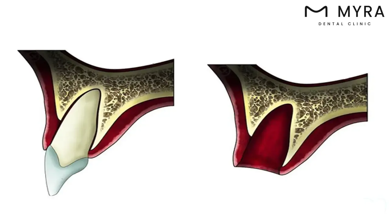
The maxillary central incisors are more vulnerable to damage and decay because of their conspicuous position in the front of the mouth, which results in inflammation or infection of the pulp. The shallow roots of maxillary central incisors render them more vulnerable to injury or infection. The treatment allows dentists to save vital teeth from being extracted while reducing patient pain and discomfort by treating maxillary central incisors with root canal therapy. Root canal therapy facilitates maintaining the smile's integrity and beauty, which preserves the natural structure of the maxillary central incisors and promotes general oral health and well-being.
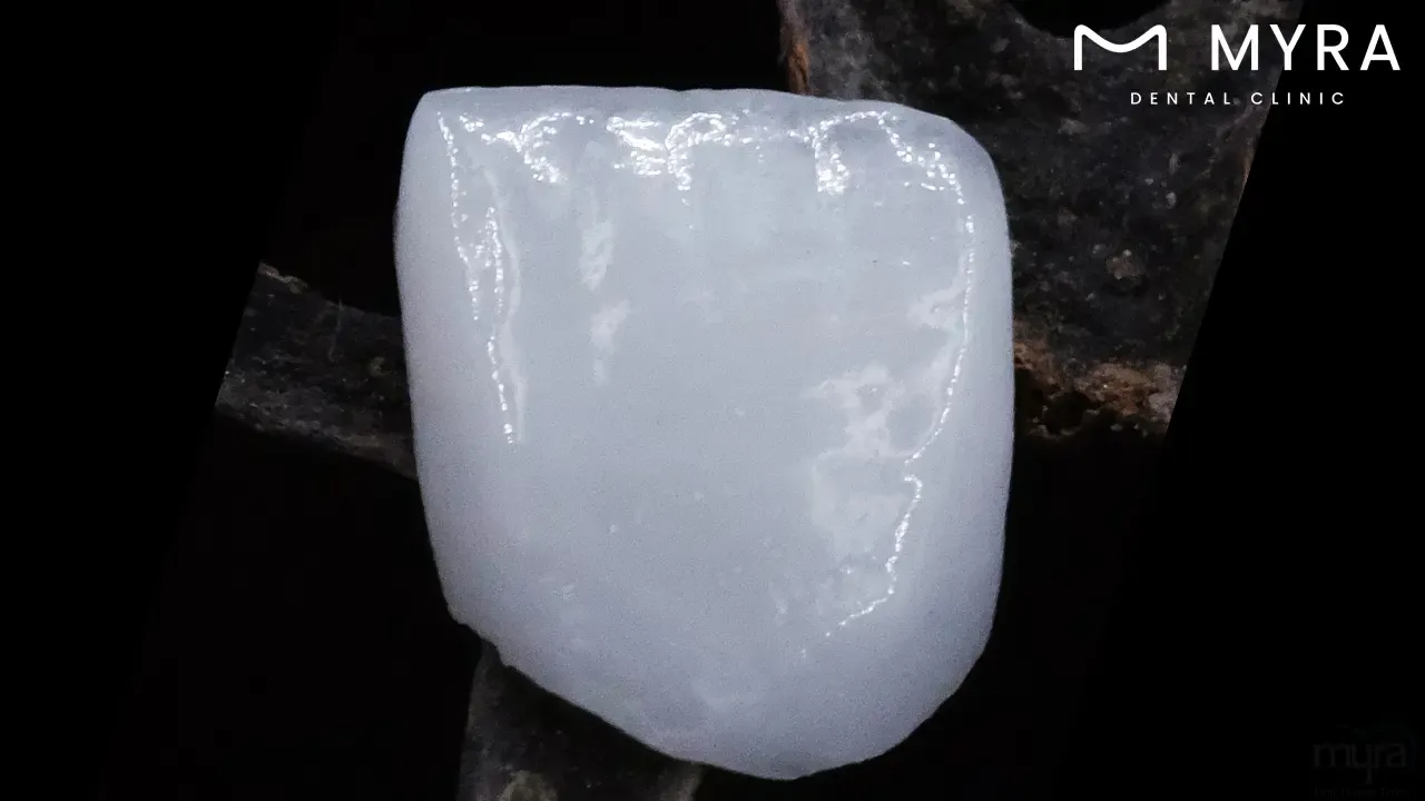
3. Dental Crowns
Dental crowns, sometimes referred to as caps, are prosthetics that are used to repair teeth that have been compromised by disease, trauma, or other causes. They are used to restore the size, strength, form, and appearance of the tooth. The standard dental operation involves applying a custom-made crown to the entire visible section of the tooth above the gumline, thus encasing and conserving the remaining tooth structure. Dental crowns are often advised when a filling or other conservative therapy is insufficient to restore a tooth effectively. The dentist uses local anaesthesia to numb the damaged tooth and surrounding area to make the patient comfortable during the crown placement process.
The dentist shapes the tooth to fit the crown and removes any decayed or damaged tissue. The prepped tooth is then imprinted and manufactured to resemble the original tooth in terms of size, shape, and colour. The prepared tooth is covered with a temporary crown to protect it while it is being worked on. Dental cement is used to securely glue the permanent crown to the tooth after the temporary crown is removed when the permanent crown is ready. The crown is polished and adjusted to ensure it fits and aligns correctly with the neighbouring teeth.
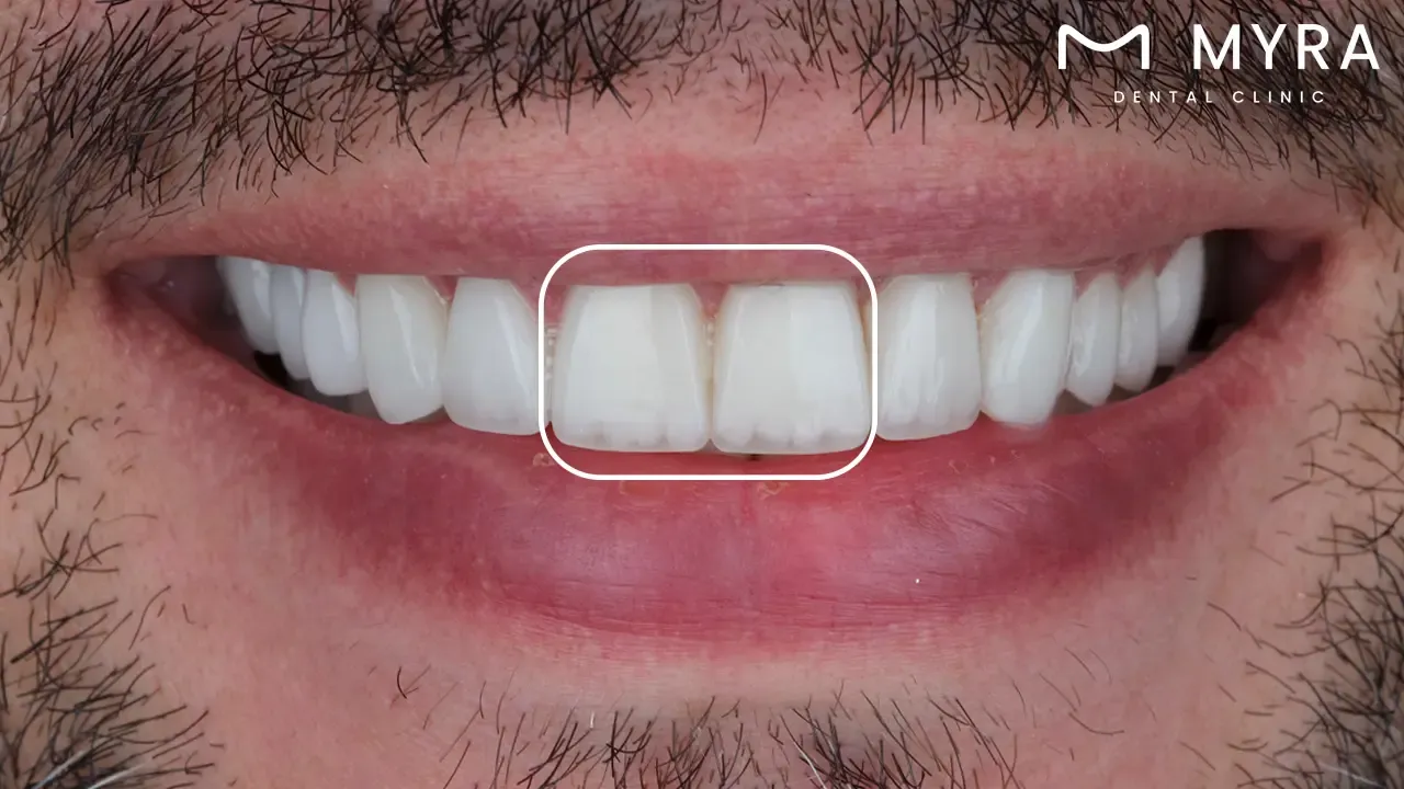
Dental crowns are a popular operation for maxillary central incisors because they are beneficial in terms of aesthetics and functionality. The maxillary central incisors are the most noticeable teeth in the upper jaw, and how they look has a big influence on how the smile appears. Maxillary central incisor teeth are often fixed with dental crowns because they are positioned firmly at the front of the mouth. It makes them extremely noticeable when smiling and vulnerable to disease or injury.
Crowns are an excellent way to restore the strength, function, and aesthetics of maxillary central incisors that have been damaged by significant decay, fractures, or other dental problems. Dentists maintain the natural structure of the significant teeth while improving their beauty, guaranteeing their long-term durability, and providing long-term functionality by crowning maxillary central incisors. Dental crowns are a crucial kind of treatment for treating a variety of dental issues and preserving the appearance and health of the maxillary central incisors.
4. Dental Veneers
Dental veneers consist of thin, custom-made shells to fit over the front surface of teeth to give a more attractive appearance. They are commonly constructed of porcelain or composite resin. The cosmetic dentistry procedure is mostly used to treat aesthetic issues, involving tooth discoloration, chipping, cracks, gaps, or misalignment, and needs minimal alterations of the teeth. The dentist first prepares the teeth's surface by removing a tiny bit of enamel to make room for the thickness of the veneers when placing dental veneers.
A unique dental glue is used by the dentist to attach the veneers to the teeth, resulting in a long-lasting, realistic-looking result. The veneers are tweaked and polished to create the ideal shape and look. Maxillary central incisor teeth are frequently treated with dental veneers because they are quite noticeable when smiling and frequently have aesthetic issues, including discoloration, chipping, or minor misalignment. Veneers provide a highly effective, painless way to improve the cosmetics of the smile and especially the appearance of the maxillary central incisors.
Maxillary central incisors often get dental veneers because of their central position in the smile and their great influence on the whole look. The maxillary central incisors, being the most conspicuous teeth in the upper jaw, are vital to the smile's look. Dental veneers allow dentists to alter the shape, colour, and alignment of the teeth, giving patients a brighter, more attractive, and self-assured smile. Dental veneers are long-lasting and stain-resistant, offering outcomes that improve the condition of the patient and self-esteem. Dental veneers are a common cosmetic dental operation for maxillary central incisors, serving as a conservative and aesthetically acceptable solution for correcting a variety of dental flaws and creating a beautiful smile.
5. Orthodontic Treatment
The goal of orthodontic therapy is to straighten teeth and jaws that are misaligned, crowded, or malocclusions caused by using dental tools, such as braces or clear aligners. The goal of the extensive dental operation is to establish optimal tooth alignment and occlusion, which enhances the smile's aesthetics and functionality. An extensive examination and assessment of the patient's dental and facial structures, including X-rays, pictures, and tooth imprints, often precedes the orthodontic rehabilitation process. The orthodontist creates a personalised treatment plan based on the diagnosis, considering the patient's unique requirements and objectives.
Braces are commonly used to gently shift teeth into desirable alignments over time. They are made up of brackets, wires, and bands. Invisalign and other clear aligners are a more discrete and removable option to traditional braces. They work by gradually realigning teeth through a set of custom-made aligners. Patients receive regular check-ups from their orthodontist during treatment to ensure the teeth are progressing according to schedule and to make any required modifications to the treatment plan. Maxillary central incisors are commonly treated with orthodontics because they play an important role in the alignment and appearance of the smile.
Orthodontic treatment for maxillary central incisors helps address underlying skeletal abnormalities, such as vertical maxillary excess, to create the best face appearance and harmony. Orthodontic therapy is a frequent and beneficial method for achieving a healthy, attractive smile and enhancing overall dental health and well-being, given the significance of the maxillary central incisors in the appearance and function of the smile.
A misalignment or malocclusion of the maxillary central incisors triggers functional problems such as difficulties speaking or chewing and impairs the smile's visual appeal. Orthodontists boost a patient's oral health and quality of life by using orthodontic therapy to realign the maxillary central incisors. It improves the function and appearance of the smile. Orthodontic therapy aims to avoid future dental problems, such as tooth decay, gum disease, and temporomandibular joint (TMJ) disorders, by promoting optimal tooth alignment and occlusion. Orthodontic therapy is a beneficial and widely utilised procedure for maxillary central incisors, allowing patients to obtain a healthier, more appealing smile.
6. Dental Implants
Dental implants are a common dental surgery whereby prosthetic tooth roots are surgically implanted to replace lost teeth. A dental implant is used to replace a missing maxillary central incisor that was lost due to trauma, decay, or extraction. It preserves the smile's natural alignment and aesthetic while regaining the ability to eat and speak clearly. The first step is to perform a thorough physical examination and assessment of the patient's medical and dental history, including the state of the nearby tissues and jawbone. A titanium implant fixture is surgically inserted into the mandible during the initial surgical surgery. It undergoes a process known as osseointegration in which it connects with the surrounding bone tissue. It gives the replacement tooth or teeth a solid and long-lasting base.
An abutment is fastened to the implant fixture to act as a link between the implant and the replacement tooth following a healing phase in which the implant fuses with the bone. The lost tooth or teeth are restored in terms of appearance, stability, and functionality with the fabrication and attachment of a custom-made dental crown, bridge, or denture to the abutment. Maxillary central incisors are extremely noticeable when smiling and are important for both cosmetic and practical reasons, hence, dental implants are a typical operation for them.
Dental implants contribute to the strength of the jawbone by encouraging bone growth and avoiding bone loss, which is common when teeth are absent. It keeps the neighbouring teeth in their appropriate alignment and avoids any modifications to the face structure. Dental implants are frequently seen as the ideal solution for replacing lost teeth, given the prominence and significance of the maxillary central incisors. The restoration of oral function and aesthetics leads to an enhanced quality of life.
Dental implants offer a long-term alternative for tooth replacement by providing natural-looking stability, longevity, and comfort. Dentists improve patients' well-being by restoring their confidence in their smile and general oral health by replacing a missing maxillary central incisor with a dental implant. A dental implant is a frequent and extremely effective surgery for replacing missing maxillary central incisors, providing patients with a long-term and aesthetically acceptable solution to tooth loss.
How does the maxillary central incisor position affect gummy smiles?
The maxillary central incisor position affects gummy smiles by impacting how a smile appears, especially when there is an excessive gingival display, known as a “gummy smile.” An excessive amount of gingival (tissue covering the teeth) is often visible when smiling, producing a gummy smile. Several things cause the problem, such as very thick gum tissue, a small upper lip, tense lip muscles, or irregular tooth eruption. An individual's confidence and self-esteem are impacted by an excessive show of gums as it is perceived as improperly large or ugly.
A gummy smile is a sign of underlying skeletal or dental problems, such as malocclusion or misaligned jaw, which affect oral health and function. Addressing the location of the maxillary central incisors with orthodontic therapy, dental restorations, or surgical treatments is required to enhance the appearance of a gummy smile and restore smile line harmony. A gummy smile results from vertical maxillary excess, a condition in which the top jawbone is longer than usual. Dentists assist patients attain a more balanced and self-assured smile, improving their look and general well-being, by realigning the maxillary central incisors to better achieve symmetry between the teeth and gums.