Born in Stuttgart, Germany in 1979, Dr. Dt. Erdem Çetin began his education in Kırşehir and completed his schooling there, from primary school through to high school. He then successfully completed his university education at the Faculty of Dentistry at 19 Mayıs University in Samsun.
Bone graft healing stages pictures illustrate the biological process through which the jawbone regenerates and strengthens after surgical intervention. A bone graft involves placing natural or synthetic material into the jaw to stimulate new bone growth and restore structural volume.
The procedure is needed when bone loss has occurred due to periodontal disease, trauma, long-term tooth absence, or in preparation for dental implants. Sinus lifts, ridge augmentations, and implant placements require a dental bone graft to establish a stable foundation.
Healing begins with blood clot formation and inflammation control, followed by tissue granulation, early bone regeneration, maturation, and eventual full integration for months.
Observing bone graft healing stages pictures provides valuable insight into each phase. Comparing bone graft before and after results and reviewing failed dental bone graft healing pictures helps understand clinical complications. A well-integrated bone graft supports implant stability and reduces the risk of prosthetic failure.
Recognising the dental bone graft healing timeline contributes to assessing progress and identifying signs of infection, rejection, or delayed integration, supporting predictable long-term outcomes for restorative treatments involving a bone graft.
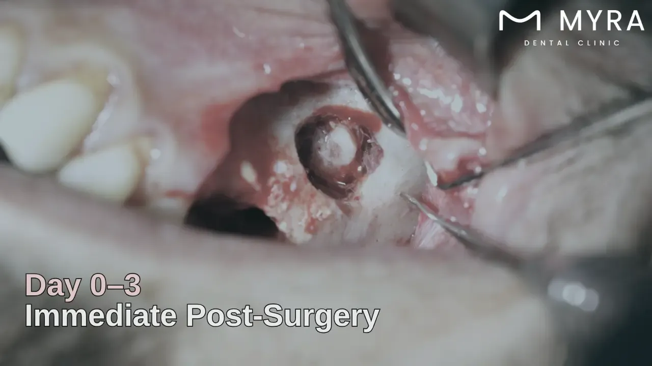
1. Immediate Post-Surgery (0-3 Days)
The immediate post-surgery phase begins after the bone graft procedure, focusing on controlling inflammation and initiating clot formation. The surgical site appears swollen, red, and slightly bruised due to soft tissue trauma and the body’s natural immune response.
A protective blood clot forms around the graft material to shield it from bacteria and physical disturbance. Discomfort and minor bleeding are expected, as healing cells concentrate at the site to prevent infection and begin early repair.
The immediate post-surgery period helps protect the graft from disturbance, allowing the stabilisation of graft particles within the surgical socket.
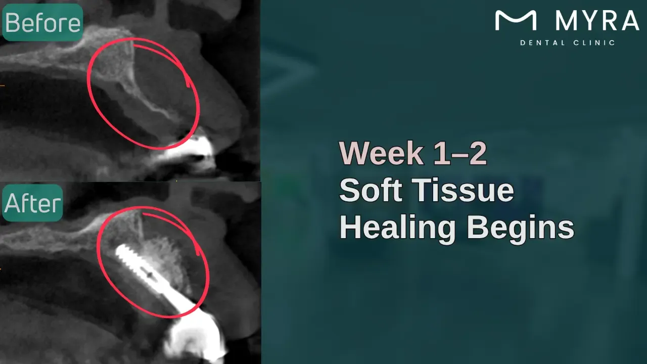
2. Initial Healing and Blood Clot Formation (1-2 Weeks)
The initial healing and blood clot formation stage marks soft tissue closing over the graft as epithelial cells regenerate across the surgical area. The blood clot formed in the initial phase matures and becomes a scaffold for new tissue growth.
Fibroblasts and endothelial cells support vascular development and collagen production, which are needed for early wound stability. Swelling reduces, and the site begins to appear uniform in colour, though tenderness remains. A layer of gum tissue covers part of the graft, promoting healing while shielding it from oral contaminants.
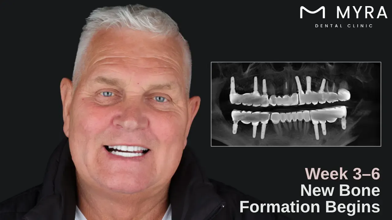
3. Early Bone Regeneration (3-6 Weeks)
The early bone regeneration stage is where the bone graft undergoes resorption and replacement, which results in the body reshaping the bone graft into living bone. Osteoblasts, the bone-forming cells, migrate into the area and deposit new bone matrix, beginning the transformation of the scaffold into a functional bone structure.
The graft integrates tightly with the surrounding jawbone, reducing the risk of displacement. Early signs of structural development are seen through radiographic imaging, even though the bone remains immature and does not have full strength. The gum tissue is fully sealed, ensuring a stable environment for uninterrupted osseous development.
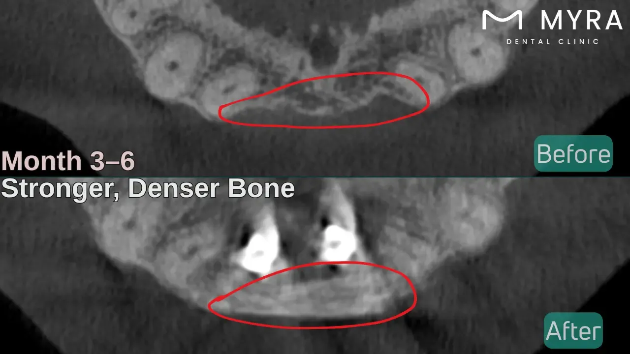
4. Bone Maturation (3-6 Months)
The bone maturation stage marks the period of mineralisation and architectural strengthening of the newly formed bone. The grafted site gradually becomes denser as immature bone is replaced with lamellar bone, which possesses higher mineral content and organised collagen fibres.
Osteoclasts contribute by removing non-viable graft particles, allowing space for mature tissue to occupy the site. The bone begins to behave like native bone, providing sufficient support for upcoming dental procedures, such as implant placement.
The affected area gains resistance against physical forces and better vascular integration, preparing for permanent prosthetic restoration.
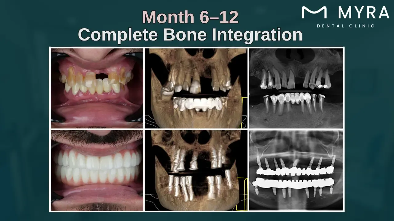
5. Full Bone Integration (6 Months-1 Year)
The full bone integration is the final healing phase, which involves full osseointegration, where the grafted bone becomes indistinguishable from the original jawbone. Cellular turnover slows as the site achieves biological stability, with mature lamellar bone exhibiting optimal density and strength. The region now supports mechanical loads required for dental implants, dentures, or crowns without structural compromise.
Radiographs show consistent bone levels, while clinical examination confirms tissue health and integration. Completing the stage results in restored jaw architecture, improved bite function, and long-term periodontal health.
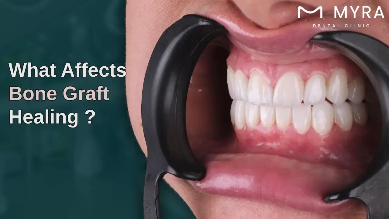
What are the Factors Affecting the Healing Stages of Dental Bone Grafts?
The Factors Affecting the Healing Stages of Dental Bone Grafts are listed below.
Type of Bone Graft: Socket preservation grafts fill extraction sites and heal faster than more complex procedures. Sinus lifting with augmentation grafts require longer healing due to the complexity and upper jaw location. Ridge expansion with augmentation grafts rebuild the alveolar ridge and need additional time to mix with the existing bone structure.
Smoking: Smoking delays healing by restricting blood flow to the surgical site. The chemicals in tobacco reduce oxygen levels in the bloodstream, which is essential for proper bone formation. Patients who smoke have a much higher risk of graft failure and complications than non-smokers.
Medical Conditions: Diabetes slows healing by impairing blood circulation and increasing infection risk through elevated blood sugar levels. Autoimmune disorders interfere with normal healing processes. The immune system reacts negatively to graft material. Osteoporosis presents challenges as it affects bone density and the body's ability to form new bone.
Nutrition: Calcium and vitamin D deficiencies impair the body's ability to create new bone tissue after grafting procedures. Protein intake supports collagen formation, which makes the framework for bone mineralisation. Patients with poor dietary habits experience delayed healing and suboptimal graft integration.
Infection: Bacterial contamination triggers inflammatory responses that break down graft material. Post-operative infections necessitate additional treatments, with antibiotics or graft removal. Proper sterilisation protocols and post-operative care prevent complications.
Medications: Bisphosphonates inhibit bone remodelling and lead to compromised healing of graft sites. Corticosteroids reduce inflammation, but suppress the immune system and slow tissue regeneration. Anticoagulants increase bleeding risk in and after surgery, destabilising the graft material.
Age: Younger patients have more blood supply and cellular activity, leading to faster bone formation and integration. Elderly patients experience slower healing times due to reduced metabolic activity and a stop in stem cell populations. The body's regenerative capacity decreases with age, requiring modified expectations for healing timelines.
Oral Hygiene: Poor oral hygiene allows bacterial build-up around the surgical site, leading to infection and graft failure. Regular but gentle cleaning helps maintain the integrity of the healing tissues surrounding the graft material. Patients must follow specific post-operative cleaning instructions to protect the graft site.
Surgical Technique: Improper placement or insufficient graft material stabilisation leads to micromotion, disrupting healing. Excessive surgical trauma damages the surrounding tissues and blood vessels that are essential for nutrient delivery. Meticulous surgical technique with minimal tissue disruption promotes optimal conditions for successful graft integration.
Post-operative Care: Following the dentist's instructions regarding physical activity and diet helps protect the graft in the initial healing phases. Avoiding pressure or trauma to the surgical site prevents graft material displacement and blood clot disruption. Attending all follow-up appointments allows for identifying and managing any healing complications.
Can Cheaper Bone Grafts still Heal Properly?
Yes, cheaper bone grafts can heal properly through the patient's natural bone tissue. Lower-cost graft materials like xenografts or animal-derived or alloplastic materials take longer to mix than premium autografts or the patient's bone. Cheaper grafts provide structural support for dental implants.
Proper surgical technique, patient health factors, and post-operative care affect success rather than the material's price point. Dental practices offer pricing options without compromising clinical outcomes, allowing patients to make informed decisions based on their budget constraints.
Research indicates that synthetic and processed graft materials achieve comparable long-term success rates to more expensive options in suitable candidates. Patients must discuss available options with their dental professional to determine the most appropriate treatment plan, considering their specific clinical needs and Bone Grafting Cost considerations, rather than assuming higher prices guarantee better results.
What are the Signs of Bone Graft Complications?
The Signs of Bone Graft Complications are listed below.
Persistent Pain and Swelling: Discomfort after surgery is normal, but pain that intensifies or persists beyond 7-10 days shows a problem. Excessive swelling, if it increases rather than decreases after the first few days, suggests infection or inflammatory response. The symptoms must be reported to the dentist when accompanied by heat or redness in the area.
Graft Material Exposure: Small particles of graft material visible through the gum tissue or actively coming out of the surgical site indicate compromised healing. The exposure creates pathways for bacteria to enter the graft site, leading to infection. Patients must avoid manipulating the area and contact their dentist if they notice any material displacement.
Fever and General Malaise: A temperature above 38°C or 100.4°F signals infection at the graft site. Accompanying symptoms include fatigue, chills, or a feeling of unwellness that develops within days after the procedure. The signs require medical evaluation, which shows an infection that needs antibiotics.
Unusual Discharge: Pus or abnormal fluid drainage from the surgical site shows active infection within the graft. The discharge has an unpleasant taste or odour and is accompanied by bad breath that persists despite regular oral hygiene. Unusual secretions must be documented and reported to the dental professional immediately.
Change in Bite or Mobility: Shifting in the position of adjacent teeth or changes in how the teeth come together when biting show graft movement or failure. Increased mobility of teeth near the graft site suggests bone support issues or inflammatory processes affecting stability. The changes develop gradually and require assessment to determine appropriate care.
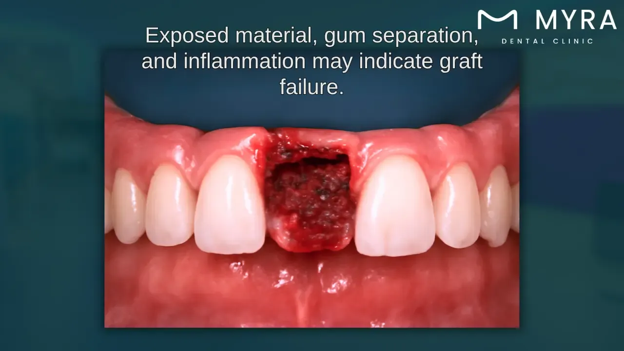
What does a Failed Bone Graft look like?
A failed bone graft looks like a raw, exposed tissue area where the graft material has not correctly integrated with the surrounding bone and soft tissue. Bone graft observes inflamed, reddened gum tissue surrounding the surgical site, with material visible through the wound.
The image shows recession and inflammation in the upper arch, with grafting material that appears white or yellowish against the red gum tissue, which shows that healing has been compromised. Failed dental bone graft healing pictures show the characteristic signs of tissue exposure. The protective gum tissue has separated from the graft site, preventing proper integration and leading to graft rejection.
What are the Best Aftercare Tips for Bone Graft Healing?
The Best Aftercare Tips for Bone Graft Healing are listed below.
Maintain Gentle Oral Hygiene: Patients must brush around the surgical site using a soft-bristled toothbrush to prevent disrupting the healing tissues. Rinsing with salt water 3-4 times daily helps keep the area clean without irritating the graft.
Follow Medication Schedule: Prescribed antibiotics must be taken exactly as directed, even if symptoms improve before finishing the course. Pain medications must be taken as the dentist recommends to manage discomfort and reduce inflammation that stops healing.
Apply Cold Compress: Ice packs must be used on the outside of the face for 15-20 minutes at a time in the first 48 hours after dental surgery. It reduces swelling and discomfort while promoting better blood circulation to the healing tissues.
Modify Diet: Patients must consume soft, lukewarm foods that require minimal chewing. Hard, crunchy, spicy, or acidic foods must be avoided to prevent damage to the surgical site or cause discomfort.
Avoid Physical Exertion: Patients must refrain from physical activities and heavy lifting for at least 3-5 days after the procedure. Excessive movement increases bleeding and pressure in the surgical area, displacing the graft material.
Elevate Head Position: Dentists recommend sleeping with the head elevated on two pillows. The position reduces blood pressure to the area and minimises swelling in the early healing phase.
Do not Disturb the Surgical Site: The area must not be touched with the tongue, fingers, or any objects that introduce bacteria or dislodge the graft material. Patients must refrain from spitting forcefully or using straws.
Stop Smoking Completely: Patients must abstain from smoking and tobacco products for at least two weeks before and after the procedure. Nicotine restricts blood flow to the gums, impeding the healing process and increasing the risk of graft failure.
Attend All Follow-up Appointments: Scheduled post-operative visits must be kept. The dentist monitors healing progress and addresses concerns early. The check-ups are essential for identifying any complications before they compromise the success of the bone graft.
Maintain Proper Hydration: Plenty of water must be consumed throughout the day to support healing and tissue regeneration. Extremely hot or cold beverages must be avoided in the first few days, as they cause discomfort or bleeding at the surgical site.
When should I see a Dentist or a Surgeon for Bone Graft Issues?
You should see a dentist or oral surgeon for Bone Graft issues immediately when you experience severe pain, excessive swelling, persistent bleeding, pus, foul odour, exposed bone, implant instability, or delayed healing. The symptoms show infection, graft rejection, or other serious complications that require prompt professional intervention.
Fever above 38°C (100.4°F) or increasing rather than decreasing pain after 3-4 days post-procedure are warning signs that must never be ignored. A professional checkup is warranted if the patient notices graft material becoming exposed through the gum tissue or if the bite feels different. Early medication by a professional means the difference between saving the graft and complete failure, requiring additional procedures.
Bone grafting is commonly performed alongside implant procedures to provide a solid and reliable foundation for dental implants. Procedures such as socket preservation, ridge augmentation, and sinus lifts are frequently used to rebuild bone that's been lost over time. Having a clear understanding of these methods and swiftly managing any complications that arise can significantly improve healing outcomes and increase the long-term success of various dental implant surgical techniques.